IRAK1 antibody (AA 301-400)
 (1 validation)
-
- Target See all IRAK1 Antibodies
- IRAK1 (Interleukin-1 Receptor-Associated Kinase 1 (IRAK1))
-
Binding Specificity
- AA 301-400
-
Reactivity
- Human, Rat
-
Host
- Rabbit
-
Clonality
- Polyclonal
-
Conjugate
- This IRAK1 antibody is un-conjugated
-
Application
- ELISA, Flow Cytometry (FACS), Immunofluorescence (Cultured Cells) (IF (cc)), Immunofluorescence (Paraffin-embedded Sections) (IF (p)), Immunohistochemistry (Paraffin-embedded Sections) (IHC (p)), Immunohistochemistry (Frozen Sections) (IHC (fro))
- Cross-Reactivity
- Human, Rat
- Predicted Reactivity
- Mouse,Dog,Cow,Pig,Chicken,Rabbit
- Purification
- Purified by Protein A.
- Immunogen
- KLH conjugated synthetic peptide derived from human IRAK1
- Isotype
- IgG
-
anti-Interleukin-1 Receptor-Associated Kinase 1 (IRAK1) (AA 530-693) antibody
IRAK1 Reactivity: Human WB, ELISA, IHC (p), IP, IF, RNAi Host: Mouse Monoclonal 3A9 unconjugated
anti-Interleukin-1 Receptor-Associated Kinase 1 (IRAK1) (AA 530-693) antibodyIRAK1 Reactivity: Human WB, ELISA, IHC (p), IP, IF Host: Mouse Monoclonal 3F7 unconjugated
anti-Interleukin-1 Receptor-Associated Kinase 1 (IRAK1) (AA 683-712), (C-Term) antibodyIRAK1 Reactivity: Human WB, IHC (p) Host: Rabbit Polyclonal RB02337 unconjugated
anti-Interleukin-1 Receptor-Associated Kinase 1 (IRAK1) (AA 530-693) antibodyIRAK1 Reactivity: Human WB, IHC (p), IP, IF, EIA Host: Mouse Monoclonal 3A9 unconjugated
anti-Interleukin-1 Receptor-Associated Kinase 1 (IRAK1) (pThr209) antibodyIRAK1 Reactivity: Human, Mouse, Rat WB, FACS, IF (cc), IHC (p) Host: Rabbit Polyclonal unconjugated
anti-Interleukin-1 Receptor-Associated Kinase 1 (IRAK1) (AA 348-381), (pSer376) antibodyIRAK1 Reactivity: Human WB, IHC (p), IF Host: Rabbit Polyclonal RB56575 unconjugated
anti-Interleukin-1 Receptor-Associated Kinase 1 (IRAK1) (AA 424-633) antibodyIRAK1 Reactivity: Human WB, IF Host: Rabbit Polyclonal unconjugated
anti-Interleukin-1 Receptor-Associated Kinase 1 (IRAK1) (AA 683-712) antibodyIRAK1 Reactivity: Human WB, ELISA, IHC Host: Rabbit Polyclonal unconjugated
anti-Interleukin-1 Receptor-Associated Kinase 1 (IRAK1) (pThr387) antibodyIRAK1 Reactivity: Human, Mouse, Rat WB, ELISA, FACS, IF (cc), IF (p), IHC (p), IHC (fro) Host: Rabbit Polyclonal unconjugated
anti-Interleukin-1 Receptor-Associated Kinase 1 (IRAK1) (pThr209) antibodyIRAK1 Reactivity: Human ELISA, IF (cc), IF (p), IHC (p), IHC (fro), ICC Host: Rabbit Polyclonal unconjugated
-
- Application Notes
-
ELISA 1:500-1000
FCM 1:20-100
IHC-P 1:200-400
IHC-F 1:100-500
IF(IHC-P) 1:50-200
IF(IHC-F) 1:50-200
IF(ICC) 1:50-200 - Restrictions
- For Research Use only
-
- by
- Alamo Laboratories Inc
- No.
- #029809
- Date
- 09/03/2014
- Antigen
- Lot Number
- 140120
- Method validated
- Western Blotting
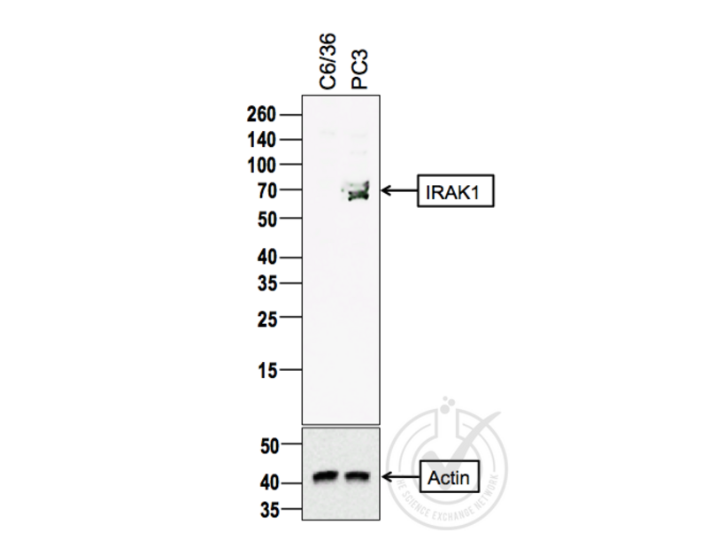
- Positive Control
- PC3 cells
- Negative Control
- C6/36 cells (non-reactive species)
- Notes
- A band was observed in the positive control sample at the correct molecular weight, which was absent from the negative control sample. Additional bands were also observed in the positive control sample, which were absent from the negative control. These bands may represent alternative IRAK1 isoforms.
- Primary Antibody
- Antigen: Interleukin-1 Receptor-Associated Kinase 1 (IRAK1)
- Catalog number: ABIN1387749
- Lot number: 140120
- Antibody Dilution: 1:100
- Secondary Antibody
- Antigen: Goat Anti-Rabbit IgG (H + L)-HRP Conjugate
- Lot number: L170-6515
- Antibody Dilution: 1:10,000
- Full Protocol
- 1. The cell extracts were heated at 95°C for 5 minutes in 1X SDS Sample Buffer containing 1% SDS and 1.25% β-mercaptoethanol.
- 2. 15 μl of heated culture-media were loaded and resolved on 8-16% SDS-polyacrylamide gel.
- 3. The Thermo Scientific - Spectra Multicolor Broad Range (Cat # 26634) were used as molecular mass markers.
- 4. Proteins were then transferred onto PVDF membrane by wet transfer and protein transfer was confirmed with Ponceau-S staining.
- 5. The PVDF membrane was incubated with 25 ml of blocking buffer [Tris Buffered Saline, pH 7.4 plus 0.1% TW20 (TBST)] containing 5% (W/V) BSA at room temperature for 1 hour.
- 6. The membrane was rinsed with TBST once.
- 7. The membrane was immersed with the protein side up in the primary antibody solution in TBST containing 5% (W/V) BSA and incubated for 24 hours at 4°C.
- 8. The membrane was rinsed in TBST thrice for 5 minutes each.
- 9. The membrane was incubated in the HRP-conjugated secondary antibody solution in TBST containing 5% (W/V) BSA and incubated for 1 hour at room temperature (~26°C) with gentle agitation.
- 10. The membrane was rinsed thrice TBST thrice for 5 minutes each.
- 11. The membrane was rinsed in TBS twice for 30 seconds each.
- 12. Signals were detected with ECL-2 Substrate. The blot was scanned for 45 minutes.
- 13. The membrane was rinsed three times TBST.
- 14. Incubated in Acidic Glycine Stripping Buffer at room temperature with gentle agitation for 3 times, 10 minutes each.
- 15. The membrane was washed in TBST 2 times for 10 minutes each.
- 16. Repeated Steps 5-12 with the loading control antibody (for Anti-actin) and its matching secondary antibody.
- Experimental Notes
- - No experimental challenges noted.
Validation #029809 (Western Blotting)
 Validation Images
![Figure 1: Western Blot for IRAK1. Arrowhead indicates the expected molecular weight of ~78 kDa.]() Figure 1: Western Blot for IRAK1. Arrowhead indicates the expected molecular weight of ~78 kDa.
Full Methods
Figure 1: Western Blot for IRAK1. Arrowhead indicates the expected molecular weight of ~78 kDa.
Full Methods -
- Format
- Liquid
- Concentration
- 1 μg/μL
- Buffer
- 0.01M TBS( pH 7.4) with 1 % BSA, 0.02 % Proclin300 and 50 % Glycerol.
- Preservative
- ProClin
- Precaution of Use
- This product contains ProClin: a POISONOUS AND HAZARDOUS SUBSTANCE, which should be handled by trained staff only.
- Storage
- 4 °C,-20 °C
- Storage Comment
- Shipped at 4°C. Store at -20°C for one year. Avoid repeated freeze/thaw cycles.
- Expiry Date
- 12 months
-
- Target
- IRAK1 (Interleukin-1 Receptor-Associated Kinase 1 (IRAK1))
- Alternative Name
- IRAK1 (IRAK1 Products)
- Synonyms
- IRAK1 antibody, IRAK antibody, pelle antibody, AA408924 antibody, IRAK-1 antibody, IRAK1-S antibody, Il1rak antibody, Plpk antibody, mPLK antibody, RGD1563841 antibody, interleukin 1 receptor associated kinase 1 antibody, interleukin-1 receptor-associated kinase 1 antibody, IRAK1 antibody, irak1 antibody, Irak1 antibody
- Background
-
Synonyms: IRAK, pelle, Interleukin-1 receptor-associated kinase 1, IRAK-1, IRAK1
Background: Serine/threonine-protein kinase that plays a critical role in initiating innate immune response against foreign pathogens. Involved in Toll-like receptor (TLR) and IL-1R signaling pathways. Is rapidly recruited by MYD88 to the receptor-signaling complex upon TLR activation. Association with MYD88 leads to IRAK1 phosphorylation by IRAK4 and subsequent autophosphorylation and kinase activation. Phosphorylates E3 ubiquitin ligases Pellino proteins (PELI1, PELI2 and PELI3) to promote pellino-mediated polyubiquitination of IRAK1. Then, the ubiquitin-binding domain of IKBKG/NEMO binds to polyubiquitinated IRAK1 bringing together the IRAK1-MAP3K7/TAK1-TRAF6 complex and the NEMO-IKKA-IKKB complex. In turn, MAP3K7/TAK1 activates IKKs (CHUK/IKKA and IKBKB/IKKB) leading to NF-kappa-B nuclear translocation and activation. Alternatively, phosphorylates TIRAP to promote its ubiquitination and subsequent degradation. Phosphorylates the interferon regulatory factor 7 (IRF7) to induce its activation and translocation to the nucleus, resulting in transcriptional activation of type I IFN genes, which drive the cell in an antiviral state. When sumoylated, translocates to the nucleus and phosphorylates STAT3.
- Gene ID
- 3654
- UniProt
- P51617
- Pathways
- NF-kappaB Signaling, TLR Signaling, Neurotrophin Signaling Pathway, Activation of Innate immune Response, Cellular Response to Molecule of Bacterial Origin, Toll-Like Receptors Cascades
-


