NRD1 antibody (Middle Region)
 (1 validation)
-
- Target See all NRD1 Antibodies
- NRD1 (Nardilysin (N-Arginine Dibasic Convertase) (NRD1))
-
Binding Specificity
- Middle Region
- Reactivity
- Human, Horse, Pig, Cow
-
Host
- Rabbit
-
Clonality
- Polyclonal
-
Conjugate
- This NRD1 antibody is un-conjugated
-
Application
- Western Blotting (WB)
- Sequence
- GSKMLSVHVV GYGKYELEED GTPSSEDSNS SCEVMQLTYL PTSPLLADCI
- Predicted Reactivity
- Cow: 93%, Horse: 86%, Human: 100%, Pig: 93%
- Characteristics
- This is a rabbit polyclonal antibody against NRD1. It was validated on Western Blot using a cell lysate as a positive control.
- Purification
- Affinity Purified
- Immunogen
- The immunogen is a synthetic peptide directed towards the middle region of human NRD1
-
anti-Nardilysin (N-Arginine Dibasic Convertase) (NRD1) (AA 32-113) antibody
NRD1 Reactivity: Human WB, ELISA, IF Host: Rabbit Polyclonal unconjugated
anti-Nardilysin (N-Arginine Dibasic Convertase) (NRD1) (AA 333-361) antibodyNRD1 Reactivity: Human WB Host: Rabbit Polyclonal RB34122 unconjugated
anti-Nardilysin (N-Arginine Dibasic Convertase) (NRD1) (AA 1058-1156) antibodyNRD1 Reactivity: Human WB, ELISA Host: Mouse Polyclonal unconjugated
anti-Nardilysin (N-Arginine Dibasic Convertase) (NRD1) (AA 1079-1128) antibodyNRD1 Reactivity: Human, Horse WB Host: Rabbit Polyclonal unconjugated
-
- Application Notes
- Optimal working dilutions should be determined experimentally by the investigator.
- Comment
-
Antigen size: 1151 AA
- Restrictions
- For Research Use only
-
- by
- AG Deeg, Lehrstuhl für Physiologie, Veterinärwissenschaftliches Department, Tierärztliche Fakultät, Ludwig-Maximilians-Universität München
- No.
- #103724
- Date
- 07/19/2019
- Antigen
- NRD1
- Lot Number
- QC27244-100203
- Method validated
- Immunohistochemistry
- Positive Control
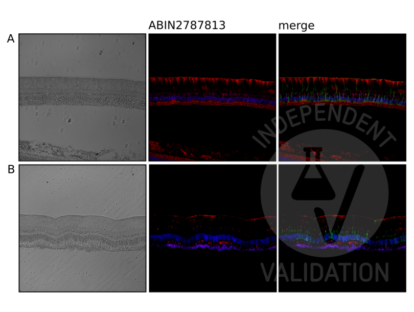
retina from horses suffering from equine recurrent uveitis (ERU)
- Negative Control
retina from non-ERU infected horses
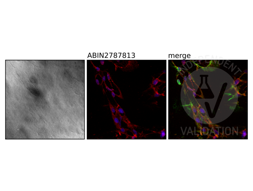
isolated equine retinal Müller glial cells (eqMCs)
- Notes
Passed. The NRD1 antibody ABIN2787813 specifically labels the targeted antigen in retinae from horses suffering from ERU and in isolated equine retinal Müller glial cells.
- Primary Antibody
- ABIN5074774
- Secondary Antibody
- goat anti-rabbit IgG (H+L) AF568-conjugated antibody (Invitrogen, A11036, lot 1832035)
- Full Protocol
- IHC on fixed equine retina:
- Dissect tissue and fix in 4% buffered formalin for 12h at 4°C.
- Dehydrate and paraffinize the tissue through a graded alcohole, xylene and paraffine series.
- Embed the tissue in Richard Allen Scientific Paraffine Type 9 (Thermofisher, 8337).
- Cut fixed tissue with a microtome into 8µm-thick sections.
- Deparaffinization and rehydration through graded xylene and graded alcohol series:
- Boil the sections in a 99°C hot waterbath in 0.1M Na2-EDTA buffer pH8.0 for 15min.
- Boil the sections in a 99°C hot waterbath in citrate buffer pH6 (Dako, 2031) for 15min.
- Rinse sections with PBS pH7.4.
- IHC on isolated eqMCs:
- Prepare eqMCs from the eyes of a 19 year old mare.
- Cultivate cells in DMEM (PAN-Biotech, P04-04510, lot 2650219) supplemented with 10% v/V FBS (Biochrom, S0615, lot 0879F) and 1% Penicillin/Streptomycin (PAN-Biotech, P06-07100, lot 7180417) for 18d at 37°C at 5% CO2.
- Seed 1x10⁵ cells (first passage) in 1ml medium on adhesion microscope slides (Langenbrinck 03-0060, lot 021218).
- Incubate cells for 2d at 37°C at 5% CO2.
- Remove medium and wash cells with PBS pH7.4.
- Fix cells for 10min with ice-cold acetone.
- Rehydrate cells with TBST (0.01M Tris, 0.15M NaCl, 0.1% v/v Tween 20) pH7.3.
- Block samples in 5% goat serum in TBST pH7.3 containing 1% w/v BSA (BSA-TBST).
- Incubate samples with primary rabbit anti-NRD1 antibody (antibodies-online, ABIN2787813, lot QC27244-100203) diluted 1:100 in BSA-TBST ON at 4°C.
- Wash samples with BSA-TBST.
- Incubate samples with secondary goat anti-rabbit IgG (H+L) AF568-conjugated antibody (Invitrogen, A11036, lot 1832035) diluted 1:500 in BSA-TBST for 30min at RT.
- Wash samples with BSA-TBST.
- Incubate samples with primary mouse anti-Vimentin antibody (Sigma-Aldrich, V6630) diluted 1:400 in BSA-TBST ON at 4°C.
- Wash samples with BSA-TBST.
- Incubate samples with secondary goat anti-mouse IgG (H+L) AF488-conjugated antibody (Invitrogen, A11029, lot 1874804) diluted 1:500 in BSA-TBST containing 0.1% DAPI for 30min at RT.
- Wash samples with BSA-TBST.
- Mount samples in Fluoromount W mounting medium (SERVA, 21634, lot 181008).
- Acquire images with Leica DMi8 fluorescence microscope equipped with Filter Cubes for DAPI, FITC, TXR and Y5 at 200x (prepared retina) and 400x (eqMCs). Parameters for image acquisition (exposure: FITC 220ms, TXR 253ms, DAPI 195ms) were maintained unchanged for all images.
- Experimental Notes
ABIN2787813 was also tested in western blot on retinal Müller glial cell lysate from healthy horses or horses suffering from ERU and in equine and porcine total retinal lysates. However, the antibody did not detect a protein of the expected MW of 132kDa in any of these samples whereas numerous extraneous protein bands were detected.
Validation #103724 (Immunohistochemistry)
 Validation Images
![IHC staining of NRD1 using ABIN2787813 (red, middle) on retina from horses suffering from equine recurrent uveitis (A) and non-ERU infected horses (B). Counterstain with DAPI (blue) and an anti-Vimentin antibody (green).]() IHC staining of NRD1 using ABIN2787813 (red, middle) on retina from horses suffering from equine recurrent uveitis (A) and non-ERU infected horses (B). Counterstain with DAPI (blue) and an anti-Vimentin antibody (green).
IHC staining of NRD1 using ABIN2787813 (red, middle) on retina from horses suffering from equine recurrent uveitis (A) and non-ERU infected horses (B). Counterstain with DAPI (blue) and an anti-Vimentin antibody (green).
![IHC staining of NRD1 using ABIN2787813 (red, middle) on isolated equine retinal Müller glial cells. Counterstain with DAPI (blue) and an anti-Vimentin antibody (green).]() IHC staining of NRD1 using ABIN2787813 (red, middle) on isolated equine retinal Müller glial cells. Counterstain with DAPI (blue) and an anti-Vimentin antibody (green).
Full Methods
IHC staining of NRD1 using ABIN2787813 (red, middle) on isolated equine retinal Müller glial cells. Counterstain with DAPI (blue) and an anti-Vimentin antibody (green).
Full Methods -
- Format
- Liquid
- Concentration
- Lot specific
- Buffer
- Liquid. Purified antibody supplied in 1x PBS buffer with 0.09 % (w/v) sodium azide and 2 % sucrose.
- Preservative
- Sodium azide
- Precaution of Use
- This product contains Sodium azide: a POISONOUS AND HAZARDOUS SUBSTANCE which should be handled by trained staff only.
- Handling Advice
- Avoid repeated freeze-thaw cycles.
- Storage
- -20 °C
- Storage Comment
- For short term use, store at 2-8°C up to 1 week. For long term storage, store at -20°C in small aliquots to prevent freeze-thaw cycles.
-
- Target
- NRD1 (Nardilysin (N-Arginine Dibasic Convertase) (NRD1))
- Alternative Name
- NRD1 (NRD1 Products)
- Synonyms
- hNRD1 antibody, hNRD2 antibody, NRDC antibody, 2600011I06Rik antibody, AI875733 antibody, NRD-C antibody, NRD1 antibody, si:dkey-171o17.4 antibody, DKFZp459N143 antibody, nardilysin convertase antibody, nardilysin, N-arginine dibasic convertase, NRD convertase 1 antibody, nardilysin antibody, nardilysin b (N-arginine dibasic convertase) antibody, NRDC antibody, Nrdc antibody, Nrd1 antibody, LOC552055 antibody, nrd1b antibody, CpipJ_CPIJ002524 antibody, Tsp_03755 antibody
- Background
-
NRD1 cleaves peptide substrates on the N-terminus of arginine residues in dibasic pairs.
Alias Symbols: hNRD1, hNRD2
Protein Interaction Partner: FBXW11, UBC, rev, SETSIP, PUS1, P3H1, ELAC2, XPO5, NSUN2, PDIA6, YARS, DNAJC7, TSN, PPP3CA, PPP2R2A, PPP2CA, PFDN5, PFDN1, PPP1R12A, MSH2, PNKP, HSPB1, MOB2, MOB1A, STK3, NRD1, HIST2H3C, UACA, COBLL1, NCOR2, NCOR1, HDAC3, IQGAP1, MAD1L1, TBL1X, UIMC1, BTR
Protein Size: 1151 - Molecular Weight
- 132 kDa
- Gene ID
- 4898
- NCBI Accession
- NM_001101662, NP_001095132
- UniProt
- O43847
- Pathways
- Skeletal Muscle Fiber Development
-


