CSF1R antibody (pTyr723)
-
- Target See all CSF1R Antibodies
- CSF1R (Colony Stimulating Factor 1 Receptor (CSF1R))
-
Binding Specificity
- pTyr723
-
Reactivity
- Human, Dog
-
Host
- Rabbit
-
Clonality
- Polyclonal
-
Conjugate
- This CSF1R antibody is un-conjugated
-
Application
- Western Blotting (WB), ELISA, Flow Cytometry (FACS), Immunohistochemistry (Paraffin-embedded Sections) (IHC (p)), Immunofluorescence (Paraffin-embedded Sections) (IF (p)), Immunofluorescence (Cultured Cells) (IF (cc)), Immunohistochemistry (Frozen Sections) (IHC (fro))
- Cross-Reactivity
- Dog, Human
- Predicted Reactivity
- Mouse,Rat,Cow,Horse
- Purification
- Purified by Protein A.
- Immunogen
- KLH conjugated synthetic phosphopeptide derived from human CSF1R around the phosphorylation site of tyrosine 723
- Isotype
- IgG
- Top Product
- Discover our top product CSF1R Primary Antibody
-
-
- Application Notes
-
WB 1:300-5000
ELISA 1:500-1000
FCM 1:20-100
IHC-P 1:200-400
IHC-F 1:100-500
IF(IHC-P) 1:50-200
IF(IHC-F) 1:50-200
IF(ICC) 1:50-200 - Restrictions
- For Research Use only
-
- by
- Sveriges Lantbruksuniversitet
- No.
- #100091
- Date
- 11/24/2016
- Antigen
- CSF1R (Colony Stimulating Factor 1 Receptor)
- Lot Number
- 9I02V3
- Method validated
- Immunohistochemistry
- Positive Control
- canine lymph node tissue
- Negative Control
- secondary antibody only
- Notes
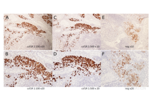
- The CSF1R antibody ABIN683788 specifically labels the targeted antigen in canine lymph node samples in IHC.
- Primary Antibody
- ABIN683788
- Secondary Antibody
- anti-rabbit (Vector, Vectastain ABC-Elite kit, PK 6101, lot 300712)
- Full Protocol
- Canine lymph node tissue is dissected and fixed in 4% buffered formalin ON at RT.
- Deparaffinization and rehydration through graded xylene and graded alcohol series:
- xylene twice for 3min.
- 100% ethanol for 1min.
- 95% ethanol for 1min.
- 70% ethanol for 1min.
- 50% ethanol for 1min.
- H2O for 2min.
- Boil the sections in the decloaking chamber (Biocare Medical):
- fill the outer chamber with 500ml distilled water.
- fill a plastic jar with diluted Antigen unmasking solution (Vector, H3301) diluted 1:100.
- Boil sections for 8 minutes, turn it off and leave for 1h.
- Rinse sections with distilled water 3 times.
- Rinse sections with 0.05M Tris-HCl pH7.6, 3 times.
- Block sections in normal goat serum (Vector, Vectastain ABC-Elite kit, PK 6101, lot 300712) diluted 1:50 for 1h at RT.
- Blot excess serum from sections; do not rinse.
- Incubate sections with primary antibody rabbit-anti CSF1R antibody (antibodies-online, ABIN683788, lot 9I02V3) diluted 1:100-1:500 in with 0.05M Tris-HCl, pH7.6 ON at 4°C. Include no primary antibody negative controls.
- Rinse sections with 0.05M Tris-HCl pH7.6, 3 times. Keep negative controls in a separate jar.
- Incubate sections with biotinylated secondary antibody anti-rabbit (Vector, Vectastain ABC-Elite kit, PK 6101, lot 300712) diluted 1:200 for 30min at RT.
- Mix 20µl of reagent A and reagent B of the Vectastain Elite ABC HRP kit (Vectorlabs, PK-6101, lot 300712) in 1ml 0.05M TrisHCl pH7.6 and leave the mixture for 30min at RT.
- Rinse sections with 0.05M Tris-HCl pH7.6, 3 times.
- Incubate sections in the ABC reagent mix for 45min at RT.
- Rinse sections with 0.05M Tris-HCl pH7.6 3 times.
- DAB staining using peroxidase substrate kit (Vector, product SK-4100, lot 020612):
- Mix thoroughly 2.5ml and 1 drop buffer stock.
- Add 2 drops DAB and mix thoroughly.
- Add 1 drop H2O2 and mix thoroughly.
- Rinse sections with 0.05M Tris-HCl pH7.6 3 times.
- Stain sections with Mayer HTX (Histolab, 01820, lot 00811.) for 2-3min at RT.
- Rinse sections in tap water for 5-10min.
- Dehydrate in fresh ethanol:
- 95% ethanol twice for 2min.
- 99.5% ethanol twice for 2min.
- xylene twice for 2min.
- Mount sections in Pertex mounting medium (Histolab, 00811).
- Dry sections in a ventilated place.
- Image acquisition (x10 and x20 objectives).
- Experimental Notes
- The blocking step with avidin and biotin was omitted for the no primary antibody negative controls. As a result, there is some unspecific staining in these sections. ABIN683788 worked nicely on the canine lymph node that was used as a positive control.
Validation #100091 (Immunohistochemistry)![Successfully validated 'Independent Validation' Badge]()
![Successfully validated 'Independent Validation' Badge]() Validation Images
Validation Images![Staining of canine lymph node tissue with ABIN683788. The primary antibody ABIN683788 was used at a 100 fold (A and B) and 500 fold (C and D) dilution. A negative control without primary antibody (E and F) was used to verify the specificity of the stain. Please refer to the protocol for detailed information. Images in the upper row (A, C, and E) were captured at 10x magnification, while the images below (B, D, and F) were taken at 20x magnification.]() Staining of canine lymph node tissue with ABIN683788. The primary antibody ABIN683788 was used at a 100 fold (A and B) and 500 fold (C and D) dilution. A negative control without primary antibody (E and F) was used to verify the specificity of the stain. Please refer to the protocol for detailed information. Images in the upper row (A, C, and E) were captured at 10x magnification, while the images below (B, D, and F) were taken at 20x magnification.
Full Methods
Staining of canine lymph node tissue with ABIN683788. The primary antibody ABIN683788 was used at a 100 fold (A and B) and 500 fold (C and D) dilution. A negative control without primary antibody (E and F) was used to verify the specificity of the stain. Please refer to the protocol for detailed information. Images in the upper row (A, C, and E) were captured at 10x magnification, while the images below (B, D, and F) were taken at 20x magnification.
Full Methods -
- Format
- Liquid
- Concentration
- 1 μg/μL
- Buffer
- 0.01M TBS( pH 7.4) with 1 % BSA, 0.02 % Proclin300 and 50 % Glycerol.
- Preservative
- ProClin
- Precaution of Use
- This product contains ProClin: a POISONOUS AND HAZARDOUS SUBSTANCE, which should be handled by trained staff only.
- Storage
- 4 °C,-20 °C
- Storage Comment
- Shipped at 4°C. Store at -20°C for one year. Avoid repeated freeze/thaw cycles.
- Expiry Date
- 12 months
-
-
: "CSF-1R as an inhibitor of apoptosis and promoter of proliferation, migration and invasion of canine mammary cancer cells." in: BMC veterinary research, Vol. 9, pp. 65, (2013) (PubMed).
-
: "CSF-1R as an inhibitor of apoptosis and promoter of proliferation, migration and invasion of canine mammary cancer cells." in: BMC veterinary research, Vol. 9, pp. 65, (2013) (PubMed).
-
- Target
- CSF1R (Colony Stimulating Factor 1 Receptor (CSF1R))
- Alternative Name
- CSF1R (CSF1R Products)
- Background
-
Synonyms: FMS, CSFR, FIM2, HDLS, C-FMS, CD115, CSF-1R, M-CSF-R, Macrophage colony-stimulating factor 1 receptor, CSF-1 receptor, CSF-1-R, Proto-oncogene c-Fms, CSF1R
Background: This protein tyrosine kinase transmembrane receptor is the receptor for colony stimulating factor 1, a cytokine which controls the production, differentiation, and function of macrophages. This receptor mediates most if not all of the biological effects of this cytokine. Ligand binding activates the receptor kinase through a process of oligomerization and transphosphorylation. The encoded protein is a member of the CSF1/PDGF receptor family of tyrosine protein kinases and contains 5 immunoglobulin like C2 type domains. CD115 is expressed by cells of the monocytic lineage and by progenitor cells. Mutations in this gene have been associated with a predisposition to myeloid malignancy.
- Gene ID
- 1436
- UniProt
- P07333
- Pathways
- RTK Signaling, Inositol Metabolic Process, Cell-Cell Junction Organization
-


 (1 reference)
 (1 validation)


