EGFR antibody
 (3 references)
 (1 validation)
-
- Target See all EGFR Antibodies
- EGFR (Epidermal Growth Factor Receptor (EGFR))
-
Reactivity
- Human
-
Host
- Rabbit
-
Clonality
- Polyclonal
-
Conjugate
- This EGFR antibody is un-conjugated
-
Application
- Western Blotting (WB), ELISA, Immunoprecipitation (IP)
- Characteristics
- Concentration Definition: by Refractometry
- Immunogen
- This whole rabbit serum was prepared by repeated immunizations with a peptide synthesized using conventional technology. The sequence of the epitope maps to a region near the carboxy terminus which is identical in human, mouse and rat EGFR.
-
anti-Epidermal Growth Factor Receptor (EGFR) (AA 693-893), (Mutant) antibody
EGFR Reactivity: Human WB, ELISA, IHC, FACS Host: Mouse Monoclonal 5G9B5 unconjugated
anti-Epidermal Growth Factor Receptor (EGFR) (AA 888-1210) antibodyEGFR Reactivity: Human WB, IHC, IP, ICC Host: Mouse Monoclonal C4 unconjugated
anti-Epidermal Growth Factor Receptor (EGFR) (pSer1026) antibodyEGFR Reactivity: Human, Mouse, Rat WB, ELISA, IHC, IF, ICC Host: Rabbit Polyclonal unconjugated
anti-Epidermal Growth Factor Receptor (EGFR) (pSer695) antibodyEGFR Reactivity: Human, Mouse, Rat WB, ELISA, IHC, IF, ICC Host: Rabbit Polyclonal unconjugated
anti-Epidermal Growth Factor Receptor (EGFR) (pSer1071) antibodyEGFR Reactivity: Human, Mouse, Rat WB, ELISA, IHC, IF, ICC Host: Rabbit Polyclonal unconjugated
anti-Epidermal Growth Factor Receptor (EGFR) (AA 1163-1191), (C-Term) antibodyEGFR Reactivity: Human WB, IHC (p) Host: Mouse Monoclonal 688CT33-1-3 unconjugated
anti-Epidermal Growth Factor Receptor (EGFR) (AA 693-893) antibodyEGFR Reactivity: Human WB, ELISA, FACS Host: Mouse Monoclonal 5E10D3 unconjugated
anti-Epidermal Growth Factor Receptor (EGFR) (C-Term) antibodyEGFR Reactivity: Human, Mouse, Rat, Monkey, Pig, Cow, Chicken, Dog, Horse, Rabbit, Guinea Pig, Avian, Cat, Donkey, Goat, Hamster, Sheep WB Host: Goat Polyclonal unconjugated
anti-Epidermal Growth Factor Receptor (EGFR) (AA 960-1060) antibodyEGFR Reactivity: Human WB, IHC, IF Host: Rabbit Polyclonal unconjugated
anti-Epidermal Growth Factor Receptor (EGFR) (AA 1021-1210) antibodyEGFR Reactivity: Human WB, IF, IP Host: Rabbit Polyclonal unconjugated
-
- Application Notes
- Anti-EGFR antibody is specifically designed for ELISA, immunoblotting and immunoprecipitation. Reactivity in other assays is likely, but has not been determined. Recognition of EGFR is independent of the phosphorylation status at tyrosine 1173. No reaction is observed against ErbB-2, ErbB-3 or ErbB-4. A431 cells, keratinocytes in normal epidermis, or placenta are typically used as positive control sources. The antigen is typically localized in the cell membrane. For western blotting, good results are also achieved on PVDF membranes blocked with 5% lowfat milk diluted in TTBS for 1 hour at room temperature. Also, dilute the primary antibody and secondary in 5% lowfat milk in TTBS. Anti-EGFR can be diluted up to 1:10,000 for immunoblot depending on the cell line and the amount of EGFR in a particular lysate. For immunoprecipitation, use approximately 10 µl of the antibody. The immunoprecipitation mix should contain the antibody, 25 µl of Protein A-agarose beads and 1.0 ml of lysate (lysate contains approximately 1.0 mg of total protein). This mixture should be rotated overnight at 4°C and then washed 3 times with lysis buffer (used to prepare the lysate). The resulting bead complex is dissolved in 20-30 µl of 3X SDS-PAGE sample buffer and approximately 15 µl is loaded per lane on an 8% polyacrylamide gel.
- Restrictions
- For Research Use only
-
- by
- ADS Biosystems Inc
- No.
- #029817
- Date
- 09/18/2014
- Antigen
- Lot Number
- 19538
- Method validated
- Western Blotting
- Positive Control
- A549 cells
- Negative Control
- MCF-7 cells
- Notes
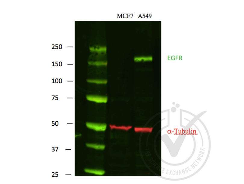
- A strong specific band was observed in the positive control at the expected size (~175 kDa) that is not observed in the negative control.
- Primary Antibody
- Antigen: Epidermal Growth Factor Receptor (EGFR)
- Catalog number: ABIN98862
- Lot number: 19538
- Dilution: 1:1,000
- Secondary Antibody
- Antibody: IRDye 680LT Goat Anti-Rabbit
- Lot number: C30725-01
- Dilution: 1:10,000
- Full Protocol
- Lysates were mixed with NuPAGE® LDS Sample Buffer (Life Technologies NP0007) and NuPAGE® Sample Reducing Agent (Life Technologies NP0004) and denatured for 5 minutes at 90ºC.
- 40 μg of each lysate was electrophoresed on a Bolt 4-12% Bis-Tris Gel (Life Technologies BG04120BOX) and run in Bolt MOPS SDS Running Buffer (Life Technologies B0001) at 160 volts for 1 hour.
- Odyssey Western Protein Standard (LI-COR #928-40000) was run as a molecular weight standard.
- PVDF membrane was activated with methanol.
- Protein samples were transferred to activated PVDF membrane in a wet Bolt Transfer Apparatus (Life Technologies B1000) at room temperature for 1 hour at 20 volts (started at 230mA, ended at 110mA).
- The membrane was blocked in x LI-COR Odyssey WB block solution for 1 hour at room temperature.
- The membrane was incubated with the primary antibody diluted 1:1000 in x LI-COR Odyssey WB block solution incubated 2 hours at room temperature.
- The membrane was washed 4 x 5 minutes in 1 x PBS-T (PBS solution with 0.1% Tween 20).
- The membrane was incubated with IRDye® 800CW Goat anti-Mouse Secondary Antibody (Red) and IRDye 680LT Goat Anti-Rabbit Secondary Antibody (Green) from LI-COR (#827-11081, Lot #C30725-01), both 1:10,000 dilutions. Incubation was performed at room temperature for 45 minutes.
- The membrane was washed 4 x 5 minutes in 1 x PBS-T (PBS solution with 0.1% Tween 20).
- Proteins were detected using Odyssey machine scanning with green channel for loading control and red channel for potential LPL band.
- Experimental Notes
- - No experimental challenges noted.
Validation #029817 (Western Blotting)
 Validation Images
![Figure 1: Scanned image of EGFR (Green) and loading control alpha-tubulin (Red) Western blot using LI-COR Odyssey Infrared Technology. First lane, protein molecular weight markers. Second lane, MCF-7 negative control lysate. Third lane, A549 positive control lysate.]() Figure 1: Scanned image of EGFR (Green) and loading control alpha-tubulin (Red) Western blot using LI-COR Odyssey Infrared Technology. First lane, protein molecular weight markers. Second lane, MCF-7 negative control lysate. Third lane, A549 positive control lysate.
Full Methods
Figure 1: Scanned image of EGFR (Green) and loading control alpha-tubulin (Red) Western blot using LI-COR Odyssey Infrared Technology. First lane, protein molecular weight markers. Second lane, MCF-7 negative control lysate. Third lane, A549 positive control lysate.
Full Methods -
- Format
- Liquid
- Concentration
- 85 mg/mL
- Preservative
- Sodium azide
- Precaution of Use
- This product contains sodium azide: a POISONOUS AND HAZARDOUS SUBSTANCE which should be handled by trained staff only.
- Storage
- -20 °C
-
-
: "Sensitivity of human granulosa cell tumor cells to epidermal growth factor receptor inhibition." in: Journal of molecular endocrinology, Vol. 52, Issue 2, pp. 223-34, (2014) (PubMed).
: "Evaluation of the cytotoxic effects of ophthalmic solutions containing benzalkonium chloride on corneal epithelium using an organotypic 3-D model." in: BMC ophthalmology, Vol. 9, pp. 5, (2009) (PubMed).
: "Downregulation of EGF receptor signaling in pancreatic islets causes diabetes due to impaired postnatal beta-cell growth." in: Diabetes, Vol. 55, Issue 12, pp. 3299-308, (2007) (PubMed).
-
: "Sensitivity of human granulosa cell tumor cells to epidermal growth factor receptor inhibition." in: Journal of molecular endocrinology, Vol. 52, Issue 2, pp. 223-34, (2014) (PubMed).
-
- Target
- EGFR (Epidermal Growth Factor Receptor (EGFR))
- Alternative Name
- EGFR (EGFR Products)
- Synonyms
- C-erb antibody, CG10079 antibody, D-EGFR antibody, D-Egf antibody, DEGFR antibody, DER antibody, DER flb antibody, DER/EGFR antibody, DER/faint little ball antibody, DER/top antibody, DER/torpedo antibody, DER1 antibody, DEgfr antibody, Degfr antibody, Der antibody, DmHD-33 antibody, Dmel\\CG10079 antibody, EFG-R antibody, EGF-R antibody, EGFR antibody, EGFr antibody, EGfr antibody, EK2-6 antibody, Egf antibody, Egf-r antibody, EgfR antibody, El antibody, Elp antibody, Elp-1 antibody, Elp-B1 antibody, Elp-B1RB1 antibody, HD-33 antibody, TOP antibody, Torpedo/DER antibody, Torpedo/Egfr antibody, c-erbB antibody, d-egf-r antibody, dEGFR antibody, dEGFR1 antibody, dEgfr antibody, der antibody, egfr antibody, flb antibody, l(2)05351 antibody, l(2)09261 antibody, l(2)57DEFa antibody, l(2)57EFa antibody, l(2)57Ea antibody, mor1 antibody, top antibody, top/DER antibody, top/flb antibody, torpedo/Egfr antibody, torpedo/egfr antibody, EGFR12 antibody, EGFR15 antibody, egfr1 antibody, Erbb2 antibody, ERBB antibody, ERBB1 antibody, HER1 antibody, PIG61 antibody, mENA antibody, ErbB-1 antibody, Errp antibody, 9030024J15Rik antibody, AI552599 antibody, Erbb antibody, Errb1 antibody, Wa5 antibody, wa-2 antibody, wa2 antibody, epidermal growth factor receptor antibody, Epidermal growth factor receptor antibody, epidermal growth factor receptor a (erythroblastic leukemia viral (v-erb-b) oncogene homolog, avian) antibody, EGFR antibody, Egfr antibody, egfra antibody, egfr1 antibody, LOC5564544 antibody
- Background
-
EGFR is a transmembrane glycoprotein that is a member of a family of protein tyrosine kinases crucial to maintaining a normal balance in cell growth and development. Growth factor receptors are involved not only in promoting the proliferation of normal cells but also in the aberrant growth of many types of human tumors. For example, the epidermal growth factor receptor (EGFR) is mutated and/or over-expressed in many common solid human squamous cell carcinomas including breast, brain, bladder, lung, gastric, head & neck, esophagus, cervix, vulva, ovary, and endometrium. Over-expression of the EGFR gene occurs in carcinomas with and without gene amplification. EGFR and ErbB-2 are particularly important in breast cancer because increased production or activation has been associated with poor prognosis. EGFR belongs to a family of growth factor receptors, which also includes ErbB-2/HER-2/neu, ErbB-3/HER-3/neu and ErbB-4/HER-4/neu. EGFR can heterodimerize with each of the members of this family.
Synonyms: Receptor tyrosine-protein kinase erbB-1 antibody, c-erbB-1 antibody - Gene ID
- 1956, 29725609
- UniProt
- P00533
- Pathways
- NF-kappaB Signaling, RTK Signaling, Fc-epsilon Receptor Signaling Pathway, EGFR Signaling Pathway, Neurotrophin Signaling Pathway, Stem Cell Maintenance, Hepatitis C, Positive Regulation of Response to DNA Damage Stimulus, Interaction of EGFR with phospholipase C-gamma, Thromboxane A2 Receptor Signaling, EGFR Downregulation, S100 Proteins
-


