Myosin VIIA antibody
 (1 validation)
-
- Target See all Myosin VIIA (MYO7A) Antibodies
- Myosin VIIA (MYO7A)
-
Reactivity
- Human, Mouse
-
Host
- Rabbit
-
Clonality
- Polyclonal
-
Conjugate
- This Myosin VIIA antibody is un-conjugated
-
Application
- ELISA, Immunohistochemistry (IHC)
- Purification
- Affinity purification
- Immunogen
- Synthetic peptide of human MYO7A
- Isotype
- IgG
-
anti-Myosin VIIA (MYO7A) (AA 838-968) antibody
MYO7A Reactivity: Human ELISA, IHC, IF Host: Rabbit Polyclonal unconjugated
anti-Myosin VIIA (MYO7A) (AA 851-950) antibodyMYO7A Reactivity: Human, Mouse ELISA, IF (cc), IF (p), IHC (fro), IHC (p) Host: Rabbit Polyclonal unconjugated
anti-Myosin VIIA (MYO7A) (AA 2118-2213) antibodyMYO7A Reactivity: Human ELISA, WB Host: Mouse Polyclonal unconjugated
anti-Myosin VIIA (MYO7A) (AA 2118-2213) antibodyMYO7A Reactivity: Human ELISA Host: Mouse Monoclonal 1D3 unconjugated
anti-Myosin VIIA (MYO7A) (AA 850-1150) antibodyMYO7A Reactivity: Human WB Host: Rabbit Polyclonal unconjugated
-
- Application Notes
- IHC 1:25-1:100
- Restrictions
- For Research Use only
-
- by
- Martinelli Lab, Neuroscience Department, UConn Health
- No.
- #103142
- Date
- 05/17/2018
- Antigen
- MYO7A
- Lot Number
- EC8591
- Method validated
- Immunofluorescence
- Positive Control
Organ of Corti tissue dissected from the cochlea of the inner ear, from a WT mouse, C57BL/6 strain, 10 weeks old.
- Negative Control
- Primary and secondary only antibody controls
- Notes
Passed. ABIN2435036 recognizes MYO7A with the expected expression pattern in murine organ of Corti tissue dissected from the cochlea of the inner ear.
- Primary Antibody
- ABIN2435036
- Secondary Antibody
- anti-rabbit AF546 conjugated antibody (Life Technologies, A11035, lot 1812311)
- Full Protocol
- Dissect organ of Corti from the mouse cochlea in the inner ear as described in Maison, Liberman, and Liberman (2016).
- Cryoprotect and freeze/thaw to permeabilize the tissue:
- Transfer cochlear pieces to a 5ml disposable cup with approximately 1ml of 30% sucrose in 100mM phosphate buffer (PBS) at RT.
- Incubate tissue on a shaker for 15min at RT.
- Wash tissue with 30% sucrose in PBS at RT.
- Incubate tissue on a shaker for 15min at RT.
- Place cup on dry ice until contents freeze completely.
- Allow cup to thaw at RT.
- Pipet out the sucrose solution and wash tissue 3x for 15min with PBS containing 0.1% triton X-100 on a shaker at RT.
- Block tissue with blocking solution (5% goat serum containing 0.3% Triton-X) on a shaker for 30-60min at RT.
- Pipet out PBS + detergent and add blocking solution.
- Cut cap off a 1.5ml microcentrifuge tube and transfer tissue pieces in the blocking solution to the flipped upside-down cap.
- Incubate tissue in the flipped upside-down caps with 100µl primary
- rabbit anti-MYO7A antibody (antibodies-online, ABIN2435036, lot EC8591) diluted 1:100 in blocking solution ON at RT on an agitator.
- chicken anti-parvalbumin (Synaptic Systems, 195006) diluted 1:400 in blocking solution ON at RT on an agitator.
- Fasten tubes onto the caps and and protect them from the light.
- Pipet out the primary antibody solution.
- Rinse tissue 3x for a total of 10min with PBS containing 0.1% Triton X-100.
- Incubate tissue in the flipped upside-down caps with 100µl secondary
- goat anti-rabbit AF546 conjugated antibody (Life Technologies, A11035, lot 1812311) diluted 1:300 in blocking solution for 1h at RT away from the light.
- goat anti-chicken AF488 conjugated antibody (Life Technologies, A11039, lot 1812246) diluted 1:300 in blocking solution for 1h at RT away from the light.
- Rinse tissue 3x for a total of 10min with PBS.
- Transfer tissue pieces onto a slide with stereocilia facing up.
- Add mounting Fluoromount-G with DAPI mounting medium (ThermoFisher Scientific, 00-4959-52, lot B2215-N915) then coverslip.
- Image acquisition on a Zeiss Axiovert epifluorescence with with a 63x objective, using a Zeiss ApoTome for optical sectioning.
- Experimental Notes
The observed signal on outer hair cells in the organ of Corti for ABIN2435036 appears as expected, compared to numerous other publications. For example, Figure 4 in He et al., and Figure 1 in Kaur et al., and Figure 4 in Li et al..
ABIN2435036 worked well enough to detect the hair cells at 1:100 dilution. A more concentrated dilution would likely improve the signal to noise ratio.
No signal was observed with either negative control.
Validation #103142 (Immunofluorescence)
 Validation Images
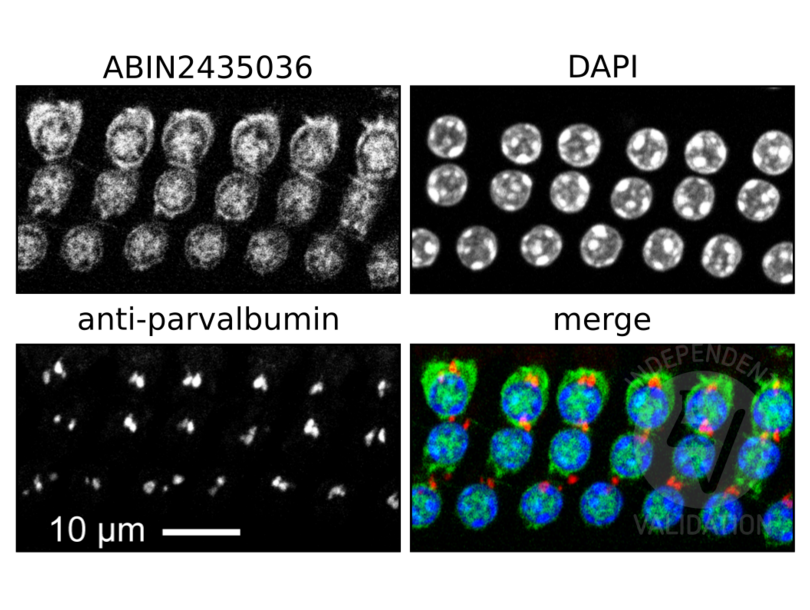
![In this photograph of the 3 rows of cochlear outer hair cells taken with 63x objective, MYO7A is labeled with ABIN2435036 in green, nuclei are labeled with DAPI (blue), and red marks the parvalbumin signal. Note that MYO7A-positive cells are adjacent to the parvalbumin-positive synapses, as expected since MYO7A is expressed in the outer hair cells. MYO7A signal can thus be used to assist in the identification and counting of outer hair cells.]() In this photograph of the 3 rows of cochlear outer hair cells taken with 63x objective, MYO7A is labeled with ABIN2435036 in green, nuclei are labeled with DAPI (blue), and red marks the parvalbumin signal. Note that MYO7A-positive cells are adjacent to the parvalbumin-positive synapses, as expected since MYO7A is expressed in the outer hair cells. MYO7A signal can thus be used to assist in the identification and counting of outer hair cells.
Full Methods
In this photograph of the 3 rows of cochlear outer hair cells taken with 63x objective, MYO7A is labeled with ABIN2435036 in green, nuclei are labeled with DAPI (blue), and red marks the parvalbumin signal. Note that MYO7A-positive cells are adjacent to the parvalbumin-positive synapses, as expected since MYO7A is expressed in the outer hair cells. MYO7A signal can thus be used to assist in the identification and counting of outer hair cells.
Full Methods -
- Format
- Liquid
- Concentration
- 0.5 mg/mL
- Buffer
- PBS with 0.05 % sodium azide and 50 % glycerol, PH7.4
- Preservative
- Sodium azide
- Handling Advice
- Avoid freeze / thaw cycles.
- Storage
- -20 °C
- Storage Comment
- Store at -20°C. Avoid freeze / thaw cycles.
-
- Target
- Myosin VIIA (MYO7A)
- Alternative Name
- MYO7A (MYO7A Products)
- Synonyms
- 53D10S antibody, BG:DS00929.11 antibody, CG7595 antibody, D antibody, D2 antibody, DM7a antibody, DMyoVIIa antibody, Dm 35B antibody, DmVIIA antibody, DmVIIa antibody, Dmel\\CG7595 antibody, Dro35B antibody, ESTS:53D10S antibody, Mhc35BC antibody, MyoVIIA antibody, anon-35Bb antibody, br27 antibody, ck/MyoVIIA antibody, l(1)35Ca antibody, l(2)07130 antibody, l(2)35Ca antibody, l(2)br27 antibody, l35Ca antibody, myoVIIA antibody, stc antibody, stch antibody, myo7a antibody, MYO7A antibody, NV17859 antibody, Hdb antibody, Myo7 antibody, USH1B antibody, nmf371 antibody, polka antibody, sh-1 antibody, sh1 antibody, DFNA11 antibody, DFNB2 antibody, MYOVIIA antibody, MYU7A antibody, NSRD2 antibody, crinkled antibody, myosin VIIAa antibody, myosin VIIa antibody, myosin VIIA antibody, myosin 7A antibody, myosin-VIIa antibody, ck antibody, myo7aa antibody, LOAG_02192 antibody, MYO7A antibody, myo7a antibody, LOC663995 antibody, LOC100116446 antibody, Myo7a antibody
- Background
- This gene is a member of the myosin gene family. Myosins are mechanochemical proteins characterized by the presence of a motor domain, an actin-binding domain, a neck domain that interacts with other proteins, and a tail domain that serves as an anchor. This gene encodes an unconventional myosin with a very short tail. Defects in this gene are associated with the mouse shaker-1 phenotype and the human Usher syndrome 1B which are characterized by deafness, reduced vestibular function, and (in human) retinal degeneration. Alternative splicing results in multiple transcript variants.
- NCBI Accession
- NP_000251
- Pathways
- Sensory Perception of Sound
-


