Histone 3 antibody (H3K36me3)
 (4 references)
 (1 validation)
-
- Target See all Histone 3 (H3) Antibodies
- Histone 3 (H3)
-
Binding Specificity
- H3K36me3
-
Reactivity
- Human, Mouse
-
Host
- Mouse
-
Clonality
- Monoclonal
-
Conjugate
- This Histone 3 antibody is un-conjugated
-
Application
- Western Blotting (WB), Chromatin Immunoprecipitation (ChIP), Dot Blot (DB), ChIP DNA-Sequencing (ChIP-seq), Cleavage Under Targets and Release Using Nuclease (CUT&RUN), Cleavage Under Targets and Tagmentation (CUT&Tag)
- Purification
- Protein G Chromatography
- Immunogen
- This Histone H3 trimethylLys36 antibody was raised against a peptide containing trimethylLys36 of human Histone H3.
- Clone
- MABI 0333
- Isotype
- IgG1
- Top Product
- Discover our top product H3 Primary Antibody
-
anti-Histone 3 (H3) (H3K4me) antibody
H3 Reactivity: Human WB, IF, ChIP, IP, ChIP-seq, CUT&RUN Host: Rabbit Polyclonal unconjugated
anti-Histone 3 (H3) (H3K27ac) antibodyH3 Reactivity: Human, Saccharomyces cerevisiae WB, IF, ChIP, DB, ICC, ChIP-seq, CUT&RUN, CUT&Tag Host: Rabbit Polyclonal unconjugated
anti-Histone 3 (H3) (3meLys4) antibodyH3 Reactivity: Human, Saccharomyces cerevisiae WB, IF, ChIP, DB, ICC, ChIP-seq, CUT&RUN Host: Rabbit Polyclonal unconjugated
anti-Histone 3 (H3) (H3K9ac) antibodyH3 Reactivity: Human, Mouse WB, IF, ChIP, DB, ICC, ChIP-seq, CUT&RUN, CUT&Tag Host: Rabbit Polyclonal unconjugated
anti-Histone 3 (H3) (AA 71-136) antibodyH3 Reactivity: Human, Mouse, Rat WB, ELISA, ICC, FACS, IHC (p), IF (cc), IF (p), IHC (fro) Host: Rabbit Polyclonal unconjugated
anti-Histone 3 (H3) (H3K27me) antibodyH3 Reactivity: Human WB, IHC, IF, ChIP, IP, ChIP-seq Host: Rabbit Polyclonal unconjugated
anti-Histone 3 (H3) (H3K4me3) antibodyH3 Reactivity: Human, Mouse, Saccharomyces cerevisiae WB, IF, ChIP, DB, ICC, ChIP-seq, CUT&RUN, CUT&Tag Host: Rabbit Polyclonal unconjugated
anti-Histone 3 (H3) (C-Term) antibodyH3 Reactivity: Human WB, IHC, ChIP, IP Host: Rabbit Polyclonal unconjugated
anti-Histone 3 (H3) (H3K9me2) antibodyH3 Reactivity: Human WB, IHC, IF, ChIP, IP, ChIP-seq Host: Rabbit Polyclonal unconjugated
anti-Histone 3 (H3) (H3K4me3) antibodyH3 Reactivity: Human WB, IHC, IF, ChIP, IP, DB, ChIP-seq, CUT&RUN, CUT&Tag Host: Rabbit Polyclonal unconjugated
-
- Application Notes
-
Recommended starting concentrations are
ChIP: 5 - 10 µg per ChIP
ChIP-Seq: 5 - 10 µg each
WB: 0.5 - 2 µg/mL dilution
DB: 0.5 - 2 µg/mL dilution
CUT&RUN: 2 µL/200 µL reaction
Optimal working dilution should be determined by the investigator. - Restrictions
- For Research Use only
-
- by
- Anna Nordin and Claudio Cantù; Cantù Lab, Gene Regulation during Development and Disease, Linköping University
- No.
- #104510
- Date
- 08/14/2023
- Antigen
- H3K36me3
- Lot Number
- 20822015
- Method validated
- Cleavage Under Targets and Release Using Nuclease
- Positive Control
Polyclonal rabbit anti-H3K4me (antibodies-online, ABIN3023251)
- Negative Control
Polyclonal guinea pig anti-rabbit IgG (antibodies-online, ABIN101961)
- Notes
Passed. ABIN2668403 allows for specific targeting of H3K36me3 in human cells using CUT&RUN.
- Primary Antibody
- ABIN2668403
- Secondary Antibody
- Full Protocol
- Cell harvest
- Harvest 50,000 human fibroblast cells per antibody.
- Centrifuge cell solution 3 min at 600 x g at RT.
- Remove the liquid carefully.
- Gently resuspend cells in 1 mL of Nuclear Extraction Buffer (20 mM HEPES-KOH pH 8.2, 20% Glycerol, 0,05% IGEPAL, 0.5 mM Spermidine, 10 mM KCl, Roche Complete Protease Inhibitor EDTA-free).
- Move the solution to a 2 mL centrifuge tube.
- Pellet the nuclei 800 x g for 5 min.
- Repeat the NE Buffer wash twice for a total of three washes.
- Resuspend the nuclei in 20 µL NE Buffer per sample.
- Concanavalin A beads preparation
- Prepare one 2 mL microcentrifuge tube.
- Gently resuspend the magnetic Concanavalin A Beads (antibodies-online, ABIN6923139).
- Pipette 10 µL Con A Beads slurry for each sample into the 1.5 mL microcentrifuge tube.
- Place the tube on a magnet stand until the fluid is clear. Remove the liquid carefully.
- Remove the microcentrifuge tube from the magnetic stand.
- Pipette 1 mL Binding Buffer (20 mM HEPES pH 7.5, 10 mM KCl, 1 mM CaCl2, 1 mM MnCl2) into each tube and resuspend ConA beads by gentle pipetting.
- Spin down the liquid from the lid with a quick pulse in a table-top centrifuge.
- Place the tubes on a magnet stand until the fluid is clear. Remove the liquid carefully.
- Remove the microcentrifuge tube from the magnetic stand.
- Repeat twice for a total of three washes.
- Gently resuspend the ConA Beads in a volume of Binding Buffer corresponding to the original volume of bead slurry, i.e. 10 µL per sample.
- Nuclei immobilization – binding to Concanavalin A beads
- Carefully vortex the nuclei suspension and add 10 µL of the Con A beads in Binding Buffer to the cell suspension for each sample.
- Close tube tightly incubates 10 min at 4 °C.
- Put the 2 mL tube on the magnet stand and when the liquid is clear remove the supernatant.
- Resuspend the beads in 1 mL of EDTA wash buffer (20 mM HEPES pH 7.5, 150 mM NaCl, 0.5 mM Spermidine, Roche Complete Protease Inhibitor EDTA-free, 2mM EDTA).
- Incubate 5 min at RT.
- Place the tube on the magnet stand and when the liquid is clear remove the supernatant.
- Resuspend the beads in 200µl of Wash Buffer (20 mM HEPES pH 7.5, 150 mM NaCl, 0.5 mM Spermidine, Roche Complete Protease Inhibitor EDTA-free) for each sample.
- Cell permeabilization and primary antibody binding
- Divide nuclei suspension into separate PCR tubes, one for each antibody (200 µL per sample).
- Add 2 µL antibody (anti-H3K36me3 antibody ABIN2668403, anti-H3K4me positive control antibody ABIN3023251, and guinea pig anti-rabbit IgG negative control antibody ABIN101961) to the respective tube, corresponding to a 1:100 dilution.
- Incubate ON at 4 °C.
- Place the tubes on a magnet stand until the fluid is clear. Remove the liquid carefully.
- Remove the microcentrifuge tubes from the magnetic stand.
- Wash with 200 µL of Wash Buffer using a multichannel pipette to accelerate the process.
- Repeat the wash five times for a total of six washes.
- pAG-MNase Binding
- Prepare a 1.5 mL microcentrifuge tube containing 200 µL of pAG mix for each sample (200 µl of wash buffer + 120 ng pAG-MNase per sample).
- Place the PCR tubes with the sample on a magnet stand until the fluid is clear. Remove the liquid carefully.
- Remove tubes from the magnetic stand.
- Resuspend the beads in 200 µL of pAG-MNase premix.
- Incubate for 30 min at 4 °C.
- Place the tubes on a magnet stand until the fluid is clear. Remove the liquid carefully.
- Remove the microcentrifuge tubes from the magnetic stand.
- Wash with 200 µL of Wash Buffer using a multichannel pipette to accelerate the process.
- Repeat the wash for a total of five washes.
- Resuspend in 200 µL of Wash Buffer.
- MNase digestion and release of pAG-MNase-antibody-chromatin complexes
- Place PCR tubes on ice and allow to chill.
- Prepare a 1.5 mL microcentrifuge tube with 51 µl of 2 mM CaCl2 mix per sample (50 µl Wash Buffer + 1 µL 100 mM CaCl2) and let it chill on ice.
- Always in ice, place the samples on the magnetic rack and when the liquid is clear remove the supernatant.
- Resuspend the samples in 50 µl of the 2 mM CaCl2 mix and incubate in ice for exactly 30 min.
- Place the sample on the magnet stand and when the liquid is clear move the supernatant in fresh collection tubes with 3µl of EDTA/EGTA 0.25M (Digestion buffer).
- Resuspend the sample in 47 µl of 1x Urea STOP Buffer (8.5 M Urea, 100 mM NaCl, 2 mM EGTA, 2 mM EDTA, 0,5% IGEPAL).
- Incubate the samples 1 h at 4 °C.
- Transfer the supernatant containing the pAG-MNase-bound digested chromatin fragments to the previously collected digestion buffer.
- DNA Clean up
- Take the Mag-Bind® TotalPure NGS beads (Omega Bio-Tek, M1378-01) from the storage and wait until they are at RT.
- Add 2x volume of beads to each sample (e.g. 100 µL of beads for 50 µL of sample).
- Incubate the beads and the sample for 15 min at RT.
- During incubation prepare fresh EtOH 80%.
- Place the PCR tubes on a magnet stand and when the liquid is clear remove the supernatant.
- Add 200 µl of fresh 80% EtOH to the sample without disturbing the beads (Important!!! Do NOT resuspend the beads or remove the tubes from the magnet stand or the sample will be lost).
- Incubate 30 sec at RT.
- Remove the EtOH from the sample.
- Repeat the wash with 80% EtOH.
- Resuspend the beads in 25 µL of 10 mM Tris.
- Incubate the sample for 2 min at RT.
- Repeat the 2x beads clean up as described before (this time with 50 µL of beads for each sample).
- Resuspend the beads + DNA in 20 µL of 10 mM Tris.
- Library preparation and sequencing
- Prepare libraries using KAPA HyperPrep Kit using KAPA Dual-Indexed adapters according to protocol.
- Sequence samples on an Illumina NextSeq 500 sequencer, using a NextSeq 500/550 High Output Kit v2.5 (75 Cycles), 36bp PE.
- Bioinformatics
- Align reads the human genome (hg38) using bowtie78 with settings -X 700 -m1 -v 3. Remove duplicate reads, and sort files using samtools. Filter mapped reads for size, keeping only reads with a fragment size at or below 120 base pairs.
- Generate bedgraph files using bedtools genomecov.
- Call peaks using SEACR version 1.3, in relaxed mode, normalizing to the negative control.
- Experimental Notes
Validation #104510 (Cleavage Under Targets and Release Using Nuclease)
 Validation Images
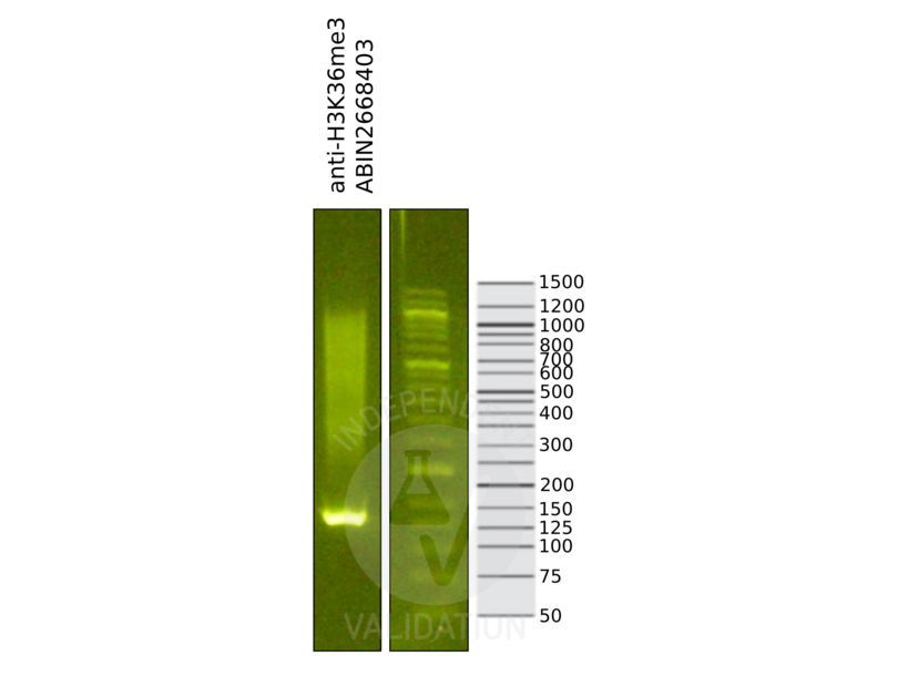
![Library profiles comparing fragment size distributions on an E-Gel EX 2% agarose gel (Thermo Fisher). Fragments obtained from CUT&RUN using anti-H3K36me3 antibody ABIN2668403 (left) after library preparation, compared to the E-Gel Sizing DNA Ladder (Thermo Fisher) (right).]() Library profiles comparing fragment size distributions on an E-Gel EX 2% agarose gel (Thermo Fisher). Fragments obtained from CUT&RUN using anti-H3K36me3 antibody ABIN2668403 (left) after library preparation, compared to the E-Gel Sizing DNA Ladder (Thermo Fisher) (right).
Library profiles comparing fragment size distributions on an E-Gel EX 2% agarose gel (Thermo Fisher). Fragments obtained from CUT&RUN using anti-H3K36me3 antibody ABIN2668403 (left) after library preparation, compared to the E-Gel Sizing DNA Ladder (Thermo Fisher) (right).
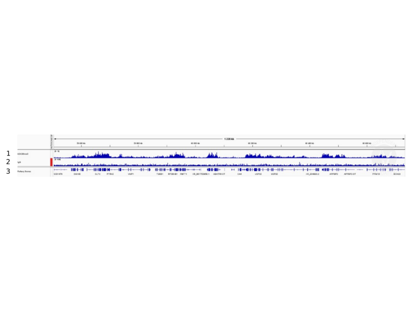
![1. Alignment tracks from CUT&RUN targeting H3K36me3 in human fibroblast cells using antibody ABIN2668403 showing the USP32 locus. 2. Alignment tracks for CUT&RUN with the IgG negative control ABIN101961. 3. RefSeq Genes.]() 1. Alignment tracks from CUT&RUN targeting H3K36me3 in human fibroblast cells using antibody ABIN2668403 showing the USP32 locus. 2. Alignment tracks for CUT&RUN with the IgG negative control ABIN101961. 3. RefSeq Genes.
Full Methods
1. Alignment tracks from CUT&RUN targeting H3K36me3 in human fibroblast cells using antibody ABIN2668403 showing the USP32 locus. 2. Alignment tracks for CUT&RUN with the IgG negative control ABIN101961. 3. RefSeq Genes.
Full Methods -
- Concentration
- 0.44 μg/μL
- Buffer
- PBS pH 7.5 containing 30 % glycerol, 0.3 M NaCl, and 0.035 % sodium azide.
- Preservative
- Sodium azide
- Precaution of Use
- This product contains Sodium azide: a POISONOUS AND HAZARDOUS SUBSTANCE which should be handled by trained staff only.
- Handling Advice
-
Avoid repeated freeze/thaw cycles and keep on ice when not in storage.
- Storage
- -20 °C
- Storage Comment
- Antibodies in solution can be stored at -20 °C for 2 years.
- Expiry Date
- 6 months
-
-
: "RSC-Associated Subnucleosomes Define MNase-Sensitive Promoters in Yeast." in: Molecular cell, Vol. 73, Issue 2, pp. 238-249.e3, (2019) (PubMed).
: "The Meiotic Recombination Activator PRDM9 Trimethylates Both H3K36 and H3K4 at Recombination Hotspots In Vivo." in: PLoS genetics, Vol. 12, Issue 6, pp. e1006146, (2016) (PubMed).
: "Arabidopsis MRG domain proteins bridge two histone modifications to elevate expression of flowering genes." in: Nucleic acids research, Vol. 42, Issue 17, pp. 10960-74, (2014) (PubMed).
: "The histone H3K36 methyltransferase MES-4 acts epigenetically to transmit the memory of germline gene expression to progeny." in: PLoS genetics, Vol. 6, Issue 9, pp. e1001091, (2010) (PubMed).
-
: "RSC-Associated Subnucleosomes Define MNase-Sensitive Promoters in Yeast." in: Molecular cell, Vol. 73, Issue 2, pp. 238-249.e3, (2019) (PubMed).
-
- Target
- Histone 3 (H3)
- Alternative Name
- Histone H3 (H3 Products)
- Synonyms
- H-3 antibody, histocompatibility 3 antibody, H3 antibody
- Molecular Weight
- 17 kDa
- Gene ID
- 3020
-


