NOTCH2 antibody
 (3 references)
 (1 validation)
Quick Overview for NOTCH2 antibody (ABIN104899)
Target
See all NOTCH2 AntibodiesReactivity
Host
Clonality
Conjugate
Application
-
-
Supplier Product No.
- 100-401-408
-
Supplier
- Rockland
-
Purpose
- NOTCH 2 Antibody
-
Cross-Reactivity (Details)
- This antiserum is directed against human NOTCH2. The immunogen epitope is only exposed after gamma secretase cleavage and is not accessible in the uncleaved form.
-
Characteristics
- Synonyms: rabbit anti-Notch2 antibody, AGS2 antibody, hN2 antibody, Notch homolog 2 antibody, neurogenic locus notch homolog protein 2
-
Purification
- Antiserum
-
Sterility
- Sterile filtered
-
Immunogen
-
Immunogen: This whole rabbit serum was prepared by repeated immunizations with a synthetic peptide corresponding to amino acid residues of human Notch 2 located near the N-terminal sequence of the cleaved N intracellular domain (NICD).
Immunogen Type: Conjugated Peptide
-
Product Specific Information
-
What can the NOTCH2 antibody ABIN104899 be used for? This polyclonal, unconjugated Rabbit anti-NOTCH2 antibody reliably detects human NOTCH2 by ELISA and Western blotting.
What validation data is available for this NOTCH2 antibody? For western blotting, independent validation is currently available and was performed at the University of Ulm, Germany. Based on the sequence, it can be assumed that the NOTCH2 antibody also works for rat and mouse species. The antibody has been used in three publications to date, which are given below and can be read at PubMed. There are 2 images available that demonstrate performance of the NOTCH2 antibody in Western blot. Use our NOTCH2 antibody to reliably detect human NOTCH 2.
What is the function of NOTCH2? NOTCH2 functions as a receptor for membrane-bound ligands Jagged-1 (JAG1), Jagged-2 (JAG2) and Delta-1 (DLL1) to regulate cell-fate determination. Upon ligand activation through the released notch intracellular domain (NICD) it forms a transcriptional activator complex with RBPJ/RBPSUH and activates genes of the enhancer of split locus. Affects the implementation of differentiation, proliferation and apoptotic programs (By similarity). NOTCH2 seems to be involved in bone remodeling and homeostasis. In collaboration with RELA/p65 enhances NFATc1 promoter activity and positively regulates RANKL-induced osteoclast differentiation. (UniProt)
-
-
anti-Notch 2 (NOTCH2) (Asp1733), (cleaved) antibody
NOTCH2 Reactivity: Human, Mouse, Rat WB, ELISA, IHC Host: Rabbit Polyclonal unconjugated
anti-Notch 2 (NOTCH2) (C-Term) antibodyNOTCH2 Reactivity: Human, Mouse WB, ChIP-seq Host: Rabbit Polyclonal unconjugated
anti-Notch 2 (NOTCH2) (AA 2251-2466) antibodyNOTCH2 Reactivity: Human WB, IHC, IP, ICC Host: Rabbit Polyclonal unconjugated
anti-Notch 2 (NOTCH2) (AA 1501-1700) antibodyNOTCH2 Reactivity: Human, Mouse WB, ELISA, IF (cc), IF (p), IHC (p), IHC (fro) Host: Rabbit Polyclonal unconjugated
anti-Notch 2 (NOTCH2) (AA 59-250) antibodyNOTCH2 Reactivity: Human ELISA, IHC, IF Host: Rabbit Polyclonal unconjugated
anti-Notch 2 (NOTCH2) (cleaved), (Internal Region) antibodyNOTCH2 Reactivity: Human WB, ELISA, IHC Host: Rabbit Polyclonal unconjugated
anti-Notch 2 (NOTCH2) (AA 2242-2471) antibodyNOTCH2 Reactivity: Human WB, IF Host: Rabbit Polyclonal unconjugated
anti-Notch 2 (NOTCH2) (AA 2421-2471) antibodyNOTCH2 Reactivity: Human WB, IP, IHC (fp) Host: Rabbit Polyclonal unconjugated
anti-Notch 2 (NOTCH2) (AA 2250-2300) antibodyNOTCH2 Reactivity: Human WB, IP, IHC (fp) Host: Rabbit Polyclonal unconjugated
anti-Notch 2 (NOTCH2) antibodyNOTCH2 Reactivity: Human, Mouse ELISA, IF, BP Host: Rabbit Chimeric B6 unconjugated Recombinant Antibody
-
-
Application Notes
-
Immunohistochemistry Dilution: 1:500
Application Note: Anti-Notch2 has been tested in ELISA, WB, and IHC. Anti-NOTCH-2 has a strong response was detected by ELISA against the immunizing peptide. This product was assayed against the peptide immunogen in a standard capture ELISA using Peroxidase conjugated anti-Rabbit IgG [H&L] (Goat) (code #611-1302) and ABTS (2,2'-azino-bis-[3-ethylbenthiazoline-6-sulfonic acid]) (code # ABTS-100) as a substrate for 30 minutes at room temperature. A working dilution of 1:30,000 to 1:90,000 is suggested in ELISA this product.
Western Blot Dilution: 1:400 - 1:2,000
ELISA Dilution: 1:20,000 - 1:100,000
Other: User Optimized
-
Restrictions
- For Research Use only
-
-
- by
- AG Pancreatic Development and Stem cell differentiation, Universitätsklinikum Ulm
- No.
- #103703
- Date
- 06/16/2019
- Antigen
- NOTCH2
- Lot Number
- Method validated
- Western Blotting
- Positive Control
pancreatic endoderm cells (day 10)
pancreatic progenitor cells (day 14)
- Negative Control
human pluripotent stem cells (day 0)
defnite endoderm cells (day 4)
- Notes
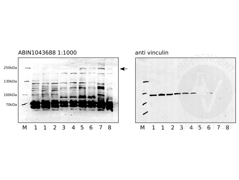
ABIN1043688 reveals two protein bands of the expected size and some weaker extraneous bands in cell lysates of human pluripotent stem cells, definite endoderm cells, pancreatic endoderm cells, and pancreatic progenitor cells.
- Primary Antibody
- ABIN1043688
- Secondary Antibody
- donkey anti-rabbit HRP-conjugated antibody (GE Healthcare, NA9310V)
- Full Protocol
- Grow HUES8 in mTeSR1 (STEMCELL Technologies, 85850) at 37°C and 5% O2, 5% CO2 in 2ml in a 6-well plate to 90% confluency.
- Harvest cells using TrypLE (ThermoFisher Scientific, 12604013) following the manufacturer´s instructions.
- Lyse 2x10⁶ cells in 30µl per well cold RIPA buffer (50mM Tris-HCl pH 8.0, 150mM NaCl, 0.1% SDS, 0.5% deoxycholate, 1% TritonX 100 in ddH2O) supplemented with 1mM PMSF and 1x protease inhibitor (Roche, 11836170001) for 30min on ice with 3x vortexing in 1.5ml microcentrifuge tubes.
- Centrifuge tubes at 10000xg for 8min at 4°C.
- Transfer supernatant to a new 1.5ml microcentrifuge tube and store at -80°C.
- Determine total protein content of the lysates using a Bradford assay (Bio-Rad, 500-0006).
- Denature 50µg of total protein for 5min at 95°C in 30µl 1x Laemmli SDS sample buffer and subsequently separate them on a denaturing 7.5% polyacrylamide gel (7.5% Acrylamide, 0.375M Tris-HCl pH8.8, 0.1% SDS, 0.1% APS, 0.1% TEMED) for about 120min at 120V.
- Transfer proteins onto a PVDF membrane (Sigma Aldrich, IPVH00010) with transfer buffer (5.27g Tris, 2.93 g glycerine, 200 ml methanol, fill to 1l ddH2O) in a semidry western blotting system for 80min at 80mA/gel.
- Check transfer of the separated proteins by Ponceau S staining.
- Rinse membrane with water.
- Wash membrane for 5min with TBST (TBS, 0.02% Tween20).
- Block the membrane with 20ml blocking buffer (TBST, 5% milk) on a shaker for 1h at RT.
- Rinse membrane 3x with TBST.
- Wash membrane on a shaker 3x for 5min with TBST.
- Shrink-wrap and incubate membrane with primary
- rabbit anti-NOTCH2 antibody (antibodies-online, ABIN1043688) diluted 1:1000 respectively in blocking buffer ON at 4°C or
- mouse anti-vinculin antibody (SigmaAldrich, V9264) diluted 1:1000 in blocking buffer at RT for 1h.
- Wash membrane 3x for 5min with TBST.
- Incubate membrane with secondary
- donkey anti-rabbit HRP-conjugated antibody (GE Healthcare, NA9310V) diluted 1:5000 in TBST containing 1% milk for 1h at RT or
- donkey anti-mouse HRP-conjugated antibody (GE Healthcare, NA9340V) diluted 1:5000 in TBST containing 1% milk for 1h at RT.
- Wash membrane 3x for 5min with TBST.
- Reveal protein bands using ECL solution (ThermoScientific, 34076) with an exposure time of 40min for the ABIN1043688 and 2min for the vinculin loading control.
- Experimental Notes
ABIN1043688 revealed some strong unspecific bands. Nevertheless, a band over 250kDa appears to be highly specific, as it is only detected at day 10 (lanes 5 and 6) and day 14 (lanes 7 and 8).
Manual exposure had to be performed due to a defect of the fusion detector in our institute.
ABIN1043688 was also tested in IHC on 4-5µm FFPE sections of primary human kidney cancer tissue. Epitope retrieval was carried out using Tris-EDTA buffer at pH9.0 (Zytomed, ZUC029-500), EDTA at pH8.0 (Leica, RE7116), or citrate buffer at pH6.1 (Agilent, S169984-2) for 20min in a decloaking chamber.
Interpretation of the results was difficult, as the method of antigen retrieval strongly effects the staining pattern. Antigen retrieval using the citrate buffer at pH6 might be ok, but knockout or knockdown studies are necessary to clearly conclusively show specificity.
Validation #103703 (Western Blotting)
 Validation Images
![Western blot with ABIN1043688 on human pluripotent stem cells (1 and 2), definite endoderm cells (3 and 4), pancreatic endoderm cells (5 and 6), and pancreatic progenitors (7 and 8). ABIN1043688 was used at a 1:1000 dilution. Vinculin served as loading control.]() Western blot with ABIN1043688 on human pluripotent stem cells (1 and 2), definite endoderm cells (3 and 4), pancreatic endoderm cells (5 and 6), and pancreatic progenitors (7 and 8). ABIN1043688 was used at a 1:1000 dilution. Vinculin served as loading control.
Full Methods
Western blot with ABIN1043688 on human pluripotent stem cells (1 and 2), definite endoderm cells (3 and 4), pancreatic endoderm cells (5 and 6), and pancreatic progenitors (7 and 8). ABIN1043688 was used at a 1:1000 dilution. Vinculin served as loading control.
Full Methods -
-
Format
- Liquid
-
Concentration
- 70 mg/mL
-
Buffer
-
Buffer: 0.02 M Potassium Phosphate, 0.15 M Sodium Chloride, pH 7.2
Stabilizer: None
Preservative: 0.01 % (w/v) Sodium Azide -
Preservative
- Sodium azide
-
Precaution of Use
- This product contains Sodium azide: a POISONOUS AND HAZARDOUS SUBSTANCE which should be handled by trained staff only.
-
Storage
- 4 °C,-20 °C
-
Storage Comment
- Store vial at -20° C prior to opening. Aliquot contents and freeze at -20° C or below for extended storage. Avoid cycles of freezing and thawing. Centrifuge product if not completely clear after standing at room temperature. This product is stable for several weeks at 4° C as an undiluted liquid. Dilute only prior to immediate use.
-
Expiry Date
- 12 months
-
-
-
: "Potassium depletion induces cellular conversion in the outer medullary collecting duct altering Notch signaling pathway." in: Scientific reports, Vol. 10, Issue 1, pp. 5708, (2020) (PubMed).
: "Notch signaling and inherited disease syndromes." in: Human molecular genetics, Vol. 12 Spec No 1, pp. R9-13, (2003) (PubMed).
: "Notch-1 and Notch-2 exhibit unique patterns of expression in human B-lineage cells." in: Leukemia, Vol. 14, Issue 12, pp. 2095-102, (2001) (PubMed).
-
-
- NOTCH2 (Notch 2 (NOTCH2))
-
Alternative Name
- NOTCH2
-
Background
- Background: Anti-Notch 2 Antibody recognizes Notch2 that is synthesized in the endoplasmic reticulum as an inactive form which is proteolytically cleaved by a furin-like convertase (S1 cleavage) in the trans-golgi network before it reaches the plasma membrane to yield an active, ligand-accessible form. Cleavage results in a C-terminal fragment N(TM) and a N-terminal fragment N(EC). Following ligand binding, it is cleaved (S2 cleavage) by TNF-alpha converting enzyme (TACE) to yield a membrane-associated intermediate fragment called Notch extracellular truncation (NEXT). This fragment is then cleaved by presenilin-dependent gamma-secretase (S3 cleavage) to release the intracellular domain (NICD) from the membrane. Anti-NOTCH2 Antibody is useful for researchers interested in Notch1, Jagged1, Jagged2, and Delta1, as well as neuroscience, transcription and cancer research.
-
Gene ID
- 4853, 24041035
-
UniProt
- Q04721
-
Pathways
- Notch Signaling, Stem Cell Maintenance
Target
-


