Iba1 antibody (C-Term)
 (13 references)
 (1 validation)
-
- Key Features
-
- High quality Iba1 Primary Antibody for the detection of IBA1.
- Reliable product with high standard validation data
- Target See all Iba1 (IBA1) Antibodies
- Iba1 (IBA1) (Ionized Calcium-binding Adapter Molecule 1 (IBA1))
-
Binding Specificity
- C-Term
-
Reactivity
- Human
-
Host
- Rabbit
-
Clonality
- Polyclonal
-
Conjugate
- This Iba1 antibody is un-conjugated
-
Application
- Western Blotting (WB), Immunohistochemistry (IHC), Immunohistochemistry (Paraffin-embedded Sections) (IHC (p)), Immunocytochemistry (ICC), Immunofluorescence (IF), Flow Cytometry (FACS), Immunohistochemistry (Frozen Sections) (IHC (fro)), Immunohistochemistry (Free Floating) (IHC (ff))
- Cross-Reactivity
- Human, Mouse, Rat
- Characteristics
-
Rabbit Polyclonal antibody to Iba1 (allograft inflammatory factor 1)
Iba1 antibody - Purification
- Purified by antigen-affinity chromatography.
- Immunogen
- Carrier-protein conjugated synthetic peptide encompassing a sequence within the C-terminus region of human Iba1. The exact sequence is proprietary.
- Isotype
- IgG
- Product Specific Information
-
What can Iba1 antibody ABIN2857032 be used for? This polyclonal rabbit anti-Iba1 antibody is unconjugated and is suitable for the detection of Iba1 / AIF1 (Allograft Inflammatory Factor 1) antigen in mouse, human, rat. It is validated for IHC and Western blotting.
What validation data is availabel for this Iba1 antibody? 18 images for IHC and immunostaining and other applications are available. The product is cited in 13 PubMed publications and has 1 independent customer validation. Use our high quality Iba1 antibody to reliably detect Iba1 / AIF1.
What is the function of Iba1 / AIF1? Iba1 is an actin-binding protein that enhances membrane ruffling and activation of RAC, increases actin-bundling activity of LCP1, binds calcium, and plays a role in RAC signaling, phagocytosis, macrophage activation and function, vascular smooth muscle cell and T-lymphocyte proliferation, lymphocyte migration, and vascular inflammation.
-
anti-Ionized Calcium-binding Adapter Molecule 1 (IBA1) (AA 51-147) antibody
IBA1 Reactivity: Human, Rat, Mouse WB, ELISA, IHC (p), FACS, IF (cc), IF (p), IHC (fro) Host: Rabbit Polyclonal unconjugated
anti-Ionized Calcium-binding Adapter Molecule 1 (IBA1) antibodyIBA1 Reactivity: Human WB, IHC, IP, IHC (p), ICC, IF, FACS, IHC (fro) Host: Rabbit Polyclonal unconjugated
anti-Ionized Calcium-binding Adapter Molecule 1 (IBA1) (AA 1-147) antibodyIBA1 Reactivity: Rat WB, IHC, IP, ICC Host: Rabbit Polyclonal unconjugated
anti-Ionized Calcium-binding Adapter Molecule 1 (IBA1) (AA 1-147) antibodyIBA1 Reactivity: Rat WB, IHC, IP, ICC Host: Mouse Monoclonal C5 unconjugated
anti-Ionized Calcium-binding Adapter Molecule 1 (IBA1) (Internal Region) antibodyVerified IBA1 Reactivity: Human IHC, ELISA, IF, FACS Host: Goat Polyclonal unconjugated
anti-Ionized Calcium-binding Adapter Molecule 1 (IBA1) (C-Term) antibodyVerified IBA1 Reactivity: Human, Rat, Mouse WB, IHC, ELISA Host: Goat Polyclonal unconjugated
anti-Ionized Calcium-binding Adapter Molecule 1 (IBA1) (AA 1-147) antibodyIBA1 Reactivity: Human WB, IHC, IF Host: Rabbit Polyclonal unconjugated
anti-Ionized Calcium-binding Adapter Molecule 1 (IBA1) antibodyIBA1 Reactivity: Mouse WB, IHC, IF Host: Rabbit Monoclonal unconjugated
anti-Ionized Calcium-binding Adapter Molecule 1 (IBA1) antibodyIBA1 Reactivity: Human WB, IHC Host: Rabbit Monoclonal unconjugated
anti-Ionized Calcium-binding Adapter Molecule 1 (IBA1) (AA 35-102) antibodyIBA1 Reactivity: Mouse WB, IHC, IP, ICC Host: Rabbit Polyclonal unconjugated
-
- Application Notes
- WB: 1:500-1:10000. ICC/IF: 1:100-1:1000. IHC-P: 1:100-1:1000. IHC-Fr: 1:100-1:1000. FACS: 1:50-1:200. Optimal dilutions/concentrations should be determined by the researcher. Not tested in other applications.
- Comment
-
Positive Control: mouse liver , rat liver
Validation: Orthogonal
- Restrictions
- For Research Use only
-
- by
- Centre d'Immunologie de Marseille-Luminy
- No.
- #101924
- Date
- 10/19/2017
- Antigen
- AIF1
- Lot Number
- 39476
- Method validated
- Immunofluorescence
- Positive Control
- PFA fixed tissue section brain of CX3CR1 GFP mouse. Staining with Iba1 (Wako, 019-19741) and Donkey anti-rabbit Fab’2 AlexaFluor 647
- Negative Control
- Notes
- Passed. Immunostaining of mouse brain samples with ABIN2857032 shows specifically the expected staining pattern.
- Primary Antibody
- ABIN2857032
- Secondary Antibody
- donkey Fab’2 anti-rabbit A647 conjugated antibody (Jackson Immunoresearch, 711-606-152, lot 128806)
- Full Protocol
- Harvest Brain from 13 weeks old mice in 1x Dulbecco’s phosphate buffered saline (DPBS) (Gibco Life Technologies, 14200-067).
- Fix mouse brain in antigen fix (Diapath, P0014) for 2h at RT.
- Wash tissue in 0.1M pH7.4 phosphate buffer at for 1h at 4°C.
- Dehydrate tissue in 30% sucrose solution ON at 4°C.
- Snap freeze tissue in Tissue Freezing Medium (ElectroMicroscopy Science, 72592-C) at -80°C.
- Cut blocks into 20µm sections using a cryostat (Leica, CM3050 S).
- Transfer sections to a slide.
- Create a hydrophobic barrier on the slide around sections with Dako pen (Dako, S2002, lot 00081640).
- Place slide in a humidified chamber and rehydrate sections in 0.1M TrisHCl pH7.4 for 10min at RT.
- Gently remove buffer by tapping slide.
- Permeabilize tissue in 0.1M TrisHCl pH7.4 containing 2% Triton X-100 (Sigma, batch 015K0039) and 0.5% BSA, for 20min at RT.
- Incubate sections with primary AIF1 (c-term) (antibodies-online, ABIN2857032, Lot 39476) diluted 1:500 in 0.1M TrisHCl pH7.4 containing 2% Triton X-100 (Sigma, batch 015K0039) and 0.5% BSA for 5h at RT. Incubate the negative contrls sections with diluted rabbit serum.
- Wash slides 1x 5min in 0.1M TrisHCl pH7.4.
- Incubate sections with secondary donkey Fab’2 anti-rabbit A647 conjugated antibody (Jackson Immunoresearch, 711-606-152, lot 128806) diluted 1:500 in 0.1M TrisHCl pH7.4 containing 2% Triton X-100 and 0.5% BSA ON at RT.
- Wash slides 1x 5min in 0.1M TrisHCl pH7.4.
- Add approximately 10μl of Slowfade® Gold antifade reagent (Life technologies, 536937, lot 1226836) for each section and mount cover slip.
- Image acquisition on a LSM 880 (Zeiss), 20x magnification, 1000 resolution.
- Experimental Notes
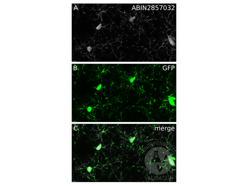
- Microglial cells in the brain of the CX3CR1 GFP mouse expressing GFP are known to express Aif-1. Immunostaining of the PFA fixed tissue sections with ABIN2857032 showed the expected co-localization of the GFP and Aif-1 signals.
Validation #101924 (Immunofluorescence)
 Validation Images
![Colocalization (C) of the signals in PFA fixed tissue sections of CX3CR1 GFP mouse brain for the immunostaining of with ABIN2857032 (A) and GFP (B).]() Colocalization (C) of the signals in PFA fixed tissue sections of CX3CR1 GFP mouse brain for the immunostaining of with ABIN2857032 (A) and GFP (B).
Full Methods
Colocalization (C) of the signals in PFA fixed tissue sections of CX3CR1 GFP mouse brain for the immunostaining of with ABIN2857032 (A) and GFP (B).
Full Methods -
- Format
- Liquid
- Concentration
- 0.06 mg/mL
- Buffer
- 1XPBS ( pH 7), 1 % BSA, 20 % Glycerol, 0.025 % ProClin 300
- Preservative
- ProClin
- Precaution of Use
- This product contains ProClin: a POISONOUS AND HAZARDOUS SUBSTANCE which should be handled by trained staff only.
- Storage
- 4 °C,-20 °C
- Storage Comment
- Store as concentrated solution. Centrifuge briefly prior to opening vial. For short-term storage (1-2 weeks), store at 4°C. For long-term storage, aliquot and store at -20°C or below. Avoid multiple freeze-thaw cycles.
-
-
: "Assessment of the Retina of Plp-α-Syn Mice as a Model for Studying Synuclein-Dependent Diseases." in: Investigative ophthalmology & visual science, Vol. 61, Issue 6, pp. 12, (2020) (PubMed).
: "Evaluation of neurological effects of cerium dioxide nanoparticles doped with different amounts of zirconium following inhalation exposure in mouse models of Alzheimer's and vascular disease." in: Neurochemistry international, Vol. 138, pp. 104755, (2020) (PubMed).
: "C-terminal binding proteins 1 and 2 in traumatic brain injury-induced inflammation and their inhibition as an approach for anti-inflammatory treatment." in: International journal of biological sciences, Vol. 16, Issue 7, pp. 1107-1120, (2020) (PubMed).
: "Lymphatic Endothelial Cells Are Essential Components of the Subcapsular Sinus Macrophage Niche." in: Immunity, Vol. 50, Issue 6, pp. 1453-1466.e4, (2019) (PubMed).
: "Multiple myeloma increases nerve growth factor and other pain-related markers through interactions with the bone microenvironment." in: Scientific reports, Vol. 9, Issue 1, pp. 14189, (2019) (PubMed).
: "Comparing the Effects of Low-Protein and High-Carbohydrate Diets and Caloric Restriction on Brain Aging in Mice." in: Cell reports, Vol. 25, Issue 8, pp. 2234-2243.e6, (2019) (PubMed).
: "NADPH oxidases as potential pharmacological targets against increased seizure susceptibility after systemic inflammation." in: Journal of neuroinflammation, Vol. 15, Issue 1, pp. 140, (2019) (PubMed).
: "Opposite microglial activation stages upon loss of PGRN or TREM2 result in reduced cerebral glucose metabolism. ..." in: EMBO molecular medicine, Vol. 11, Issue 6, (2019) (PubMed).
: "Human Umbilical Cord Mesenchymal Stem Cells Preserve Adult Newborn Neurons and Reduce Neurological Injury after Cerebral Ischemia by Reducing the Number of Hypertrophic Microglia/Macrophages." in: Cell transplantation, Vol. 26, Issue 11, pp. 1798-1810, (2018) (PubMed).
: "A scFv antibody targeting common oligomeric epitope has potential for treating several amyloidoses." in: Scientific reports, Vol. 6, pp. 36631, (2018) (PubMed).
: "Thiamine deficiency activates hypoxia inducible factor-1α to facilitate pro-apoptotic responses in mouse primary astrocytes." in: PLoS ONE, Vol. 12, Issue 10, pp. e0186707, (2017) (PubMed).
: "Astragaloside IV inhibits microglia activation via glucocorticoid receptor mediated signaling pathway." in: Scientific reports, Vol. 6, pp. 19137, (2016) (PubMed).
: "A mouse model for fucosidosis recapitulates storage pathology and neurological features of the milder form of the human disease." in: Disease models & mechanisms, Vol. 9, Issue 9, pp. 1015-28, (2016) (PubMed).
-
: "Assessment of the Retina of Plp-α-Syn Mice as a Model for Studying Synuclein-Dependent Diseases." in: Investigative ophthalmology & visual science, Vol. 61, Issue 6, pp. 12, (2020) (PubMed).
-
- Target
- Iba1 (IBA1) (Ionized Calcium-binding Adapter Molecule 1 (IBA1))
- Alternative Name
- allograft inflammatory factor 1 (IBA1 Products)
- Synonyms
- AIF-1 antibody, IBA1 antibody, IRT-1 antibody, IRT1 antibody, AI607846 antibody, D17H6S50E antibody, G1 antibody, Iba1 antibody, Bart1 antibody, iba1 antibody, mrf-1 antibody, fa04b11 antibody, wu:fa04b11 antibody, AIF antibody, AIF1 antibody, aif1 antibody, allograft inflammatory factor 1 antibody, AIF1 antibody, Aif1 antibody, aif1 antibody
- Background
-
This gene is induced by cytokines and interferon. Its protein product is thought to be involved in negative regulation of growth of vascular smooth muscle cells, which contributes to the anti-inflammatory response to vessel wall trauma. Three transcript variants encoding different isoforms have been found for this gene.
- Molecular Weight
- 17 kDa
- Gene ID
- 199
- UniProt
- P55008
- Pathways
- Smooth Muscle Cell Migration
-


