EGFR Antibodies
Your search for reliable EGFR antibodies ends here. EGFR, known by aliases such as EGFR, Egfr, egfra, egfr1, LOC5564544, is an integral part of our antibody range. Whether you're working with Human, Rat, Mouse, Cow, Pig, or other species, our range of EGFR antibodies offer precise detection across diverse samples. These specialized antibodies are tailored for various scientific applications like WB, ELISA, IHC, FACS, IF, providing you with options like polyclonal, recombinant, and monoclonal antibodies, sourced from different host species such as Rabbit, Mouse, Goat. The efficacy of our antibodies is well-established, demonstrated through multiple methods.
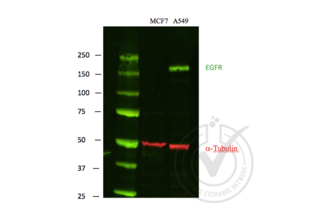
Detailed information, including references, images, and validations by other customers, can be found on each product page. Should you require assistance in finding a specific product, our customer service team is ready to assist. Utilize our EGFR antibodies in your research endeavors for dependable EGFR detection.

EGFR Reactivity: Human WB, ELISA, IHC, FACS Host: Mouse Monoclonal 5G9B5 unconjugated
EGFR Reactivity: Human WB, IHC, IP, ICC Host: Mouse Monoclonal C4 unconjugated
EGFR Antibodies by Grade
Find EGFR Antibodies with a specific Grade. The Grade listed below are among those available. Click on a link to go to the corresponding products.
EGFR Antibodies by Host
Find EGFR Antibodies with a specific Host. The Host listed below are among those available. Click on a link to go to the corresponding products.
EGFR Antibodies by Clonality
Find available monoclonal or polyclonal EGFR Antibodies. Click on a link to go to the corresponding products.
Popular EGFR Antibodies
- (3)
- (2)
- (1)
- (2)
- (7)
- (6)
- (7)
- (7)
- (8)
- (6)
- (2)
- (5)
- (6)
- (7)
- (6)
- (7)
- (5)
- (1)
- (4)
- (5)
Latest Publications for our EGFR Antibodies
: "Highly Modular Protein Micropatterning Sheds Light on the Role of Clathrin-Mediated Endocytosis for the Quantitative Analysis of Protein-Protein Interactions in Live Cells." in: Biomolecules, Vol. 10, Issue 4, (2021) (PubMed).: "Charcot-Marie-Tooth Type 2B: A New Phenotype Associated with a Novel RAB7A Mutation and Inhibited EGFR Degradation." in: Cells, Vol. 9, Issue 4, (2020) (PubMed).
: "ALIX- and ESCRT-III-dependent sorting of tetraspanins to exosomes." in: The Journal of cell biology, Vol. 219, Issue 3, (2020) (PubMed).
: "Counting growth factors in single cells with infrared quantum dots to measure discrete stimulation distributions." in: Nature communications, Vol. 10, Issue 1, pp. 909, (2019) (PubMed).
: "Age-Dependent Cellular and Behavioral Deficits Induced by Molecularly Targeted Drugs Are Reversible." in: Cancer research, Vol. 78, Issue 8, pp. 2081-2095, (2019) (PubMed).
: "Androgen receptor drives hepatocellular carcinogenesis by activating enhancer of zeste homolog 2-mediated Wnt/β-catenin signaling." in: EBioMedicine, Vol. 35, pp. 155-166, (2019) (PubMed).
: "Myosin VI and branched actin filaments mediate membrane constriction and fission of melanosomal tubule carriers." in: The Journal of cell biology, Vol. 217, Issue 8, pp. 2709-2726, (2019) (PubMed).
: "Autophagosomal YKT6 is required for fusion with lysosomes independently of syntaxin 17." in: The Journal of cell biology, Vol. 217, Issue 8, pp. 2633-2645, (2019) (PubMed).
: "Sym004-induced EGFR elimination is associated with profound anti-tumor activity in EGFRvIII patient-derived glioblastoma models." in: Journal of neuro-oncology, Vol. 138, Issue 3, pp. 489-498, (2019) (PubMed).
: "Therapeutic efficacy of a synthetic epsin mimetic peptide in glioma tumor model: uncovering multiple mechanisms beyond the VEGF-associated tumor angiogenesis." in: Journal of neuro-oncology, Vol. 138, Issue 1, pp. 17-27, (2019) (PubMed).
Aliases for EGFR Antibodies
epidermal growth factor receptor (EGFR) AntibodiesEpidermal growth factor receptor (Egfr) Antibodies
epidermal growth factor receptor a (erythroblastic leukemia viral (v-erb-b) oncogene homolog, avian) (egfra) Antibodies
epidermal growth factor receptor (egfr1) Antibodies
epidermal growth factor receptor (LOC5564544) Antibodies
epidermal growth factor receptor (Egfr) Antibodies
9030024J15Rik Antibodies
AI552599 Antibodies
C-erb Antibodies
c-erbB Antibodies
CG10079 Antibodies
D-Egf Antibodies
d-egf-r Antibodies
D-EGFR Antibodies
DEGFR Antibodies
DEgfr Antibodies
Degfr Antibodies
dEGFR Antibodies
dEgfr Antibodies
dEGFR1 Antibodies
DER Antibodies
Der Antibodies
der Antibodies
DER/EGFR Antibodies
DER/faint little ball Antibodies
DER/top Antibodies
DER/torpedo Antibodies
DER1 Antibodies
DER flb Antibodies
Dmel\\CG10079 Antibodies
DmHD-33 Antibodies
EFG-R Antibodies
Egf Antibodies
EGF-R Antibodies
Egf-r Antibodies
EGFR Antibodies
EGFr Antibodies
EGfr Antibodies
EgfR Antibodies
egfr Antibodies
egfr1 Antibodies
EGFR12 Antibodies
EGFR15 Antibodies
EK2-6 Antibodies
El Antibodies
Elp Antibodies
Elp-1 Antibodies
Elp-B1 Antibodies
Elp-B1RB1 Antibodies
ERBB Antibodies
Erbb Antibodies
ErbB-1 Antibodies
ERBB1 Antibodies
Erbb2 Antibodies
Errb1 Antibodies
Errp Antibodies
flb Antibodies
HD-33 Antibodies
HER1 Antibodies
l(2)05351 Antibodies
l(2)09261 Antibodies
l(2)57DEFa Antibodies
l(2)57Ea Antibodies
l(2)57EFa Antibodies
mENA Antibodies
mor1 Antibodies
PIG61 Antibodies
TOP Antibodies
top Antibodies
top/DER Antibodies
top/flb Antibodies
Torpedo/DER Antibodies
Torpedo/Egfr Antibodies
torpedo/Egfr Antibodies
torpedo/egfr Antibodies
wa-2 Antibodies
wa2 Antibodies
Wa5 Antibodies
Did you look for something else?
- EGFLAM Antibodies
- EGFL8 Antibodies
- EGFL7 Antibodies
- EGFL6 Antibodies
- EGF Antibodies
- EFTUD2 Antibodies
- EFTUD1 Antibodies
- EFS Antibodies
- EFR3A Antibodies
- EFNB2A Antibodies
- EFNA4 Antibodies
- EFHD2 Antibodies
- EFHD1 Antibodies
- EFHC2 Antibodies
- EFHC1 Antibodies
- EFHA1 Antibodies
- EFCAB6 Antibodies
- EFCAB4B Antibodies
- EFCAB4A Antibodies
- EFCAB1 Antibodies
- EGFR2 Antibodies
- EGLN1 Antibodies
- EGLN3 Antibodies
- EGR1 Antibodies
- EGR3 Antibodies
- EGR4 Antibodies
- EHBP1 Antibodies
- EHD1 Antibodies
- EHD2 Antibodies
- EHD3 Antibodies
- EHD4 Antibodies
- EHF Antibodies
- EHHADH Antibodies
- EHMT1 Antibodies
- EHMT2 Antibodies
- EI24 Antibodies
- EID1 Antibodies
- EID2 Antibodies
- EID2B Antibodies
- EID3 Antibodies


