CTNNB1 antibody (N-Term)
 (8 references)
 (1 validation)
-
- Key Features
-
- Rabbit beta-Catenin antibody reliably detects beta-Catenin targets in Wnt-signaling using CUT&RUN, ICC, IF, WB, ELISA.
- Well suited to image adherens junctions in IHC/IF.
- Highly specific beta-Catenin detection in WB.
- Target See all CTNNB1 Antibodies
- CTNNB1 (Catenin (Cadherin-Associated Protein), beta 1, 88kDa (CTNNB1))
-
Binding Specificity
- N-Term
-
Reactivity
- Human
-
Host
- Rabbit
-
Clonality
- Polyclonal
-
Conjugate
- This CTNNB1 antibody is un-conjugated
-
Application
- Western Blotting (WB), Immunohistochemistry (IHC), Immunofluorescence (IF), Immunohistochemistry (Paraffin-embedded Sections) (IHC (p)), Flow Cytometry (FACS), Immunoprecipitation (IP), Immunocytochemistry (ICC), Immunohistochemistry (Frozen Sections) (IHC (fro)), Chromatin Immunoprecipitation (ChIP), Cleavage Under Targets and Release Using Nuclease (CUT&RUN), Immunohistochemistry (Whole Mount) (IHC (wm))
- Cross-Reactivity
- Cat, Dog, Human, Mouse, Rabbit, Rat, Zebrafish (Danio rerio)
- Characteristics
-
Rabbit Polyclonal antibody to beta Catenin (catenin (cadherin-associated protein), beta 1, 88 kDa)
beta Catenin antibody [N1N2-2], N-term - Purification
- Purified by antigen-affinity chromatography.
- Grade
- KO Validated
- Immunogen
- Recombinant protein encompassing a sequence within the N-terminus region of human beta Catenin. The exact sequence is proprietary.
- Isotype
- IgG
- Product Specific Information
-
What can the beta-Catenin antibody ABIN2855042 be used for? This polyclonal, unconjugated rabbit anti-beta Catenin antibody is suitable for the detection of beta Catenin (CATNB, Catenin beta-1 or β-Catenin) by Chromatin Immunoprecipitation (ChIP), CUT&RUN, Immunofluorescence, ELISA, IHC and Western blotting. It has been validated for human, mouse, rat species. Additional predicted reactivities are chicken, pig, xenopus laevis, rhesus monkey and zebrafish.
What validation data is available for this beta-Catenin antibody? The beta Catenin antibody currently has an independent validation for CUT&RUN (Cleavage Under Targets and Release Using Nuclease) which can be read with protocol below. It has been used in seven publications, which are also indicated and can be found on PubMed. The rabbit beta Catenin antibody has 16 product images demonstrating its performance in CUT&RUN, Western blot, ICC / IF and IHC. Use our high-quality beta Catenin antibody to reliably detect beta Catenin for human and several other species.
What is the function of beta-Catenin? Beta Catenin is a key downstream component of the canonical Wnt signaling pathway. In the absence of Wnt, forms a complex with AXIN1, AXIN2, APC, CSNK1A1 and GSK3B that promotes phosphorylation on N-terminal Ser and Thr residues and ubiquitination of beta Catenin via BTRC and its subsequent degradation by the proteasome. In the presence of Wnt ligand, beta Catenin is not ubiquitinated and accumulates in the nucleus, where it acts as a coactivator for transcription factors of the TCF/LEF family, leading to activate Wnt responsive genes. (UniProt) It is a hard target for ChIP-seq, but you can use our beta Catenin antibody / β-Catenin antibody in CUT&RUN.
-
anti-Catenin (Cadherin-Associated Protein), beta 1, 88kDa (CTNNB1) (N-Term) antibody
CTNNB1 Reactivity: Human, Mouse, Rat WB, IHC, ELISA, IF, ICC Host: Rabbit Polyclonal unconjugated
anti-Catenin (Cadherin-Associated Protein), beta 1, 88kDa (CTNNB1) (AA 692-721), (C-Term) antibodyCTNNB1 Reactivity: Human WB, IF, IHC (p), FACS Host: Rabbit Polyclonal RB41124 unconjugated
anti-Catenin (Cadherin-Associated Protein), beta 1, 88kDa (CTNNB1) (AA 2-781) antibodyCTNNB1 Reactivity: Human WB, IHC, ELISA, IF, IP Host: Rabbit Polyclonal unconjugated
anti-Catenin (Cadherin-Associated Protein), beta 1, 88kDa (CTNNB1) (AA 78-106), (N-Term) antibodyCTNNB1 Reactivity: Human WB, IF, IHC (p), FACS Host: Rabbit Polyclonal RB41155 unconjugated
anti-Catenin (Cadherin-Associated Protein), beta 1, 88kDa (CTNNB1) (AA 1-100) antibodyCTNNB1 Reactivity: Human WB, IHC, ELISA, FACS Host: Mouse Monoclonal 7C5A2 unconjugated
anti-Catenin (Cadherin-Associated Protein), beta 1, 88kDa (CTNNB1) (AA 632-781) antibodyCTNNB1 Reactivity: Human WB, IHC, ELISA, FACS Host: Mouse Monoclonal 5B6E9 unconjugated
anti-Catenin (Cadherin-Associated Protein), beta 1, 88kDa (CTNNB1) (AA 682-781) antibodyCTNNB1 Reactivity: Human WB, ELISA, IF, PLA, RNAi Host: Mouse Monoclonal 1C9 unconjugated
anti-Catenin (Cadherin-Associated Protein), beta 1, 88kDa (CTNNB1) antibodyCTNNB1 Reactivity: Human WB, IHC, IF, FACS, StM Host: Rabbit Monoclonal CTNNB1-2030R unconjugated Recombinant Antibody
anti-Catenin (Cadherin-Associated Protein), beta 1, 88kDa (CTNNB1) antibodyCTNNB1 Reactivity: Human, Dog WB, IHC, IF, IHC (p), FACS Host: Mouse Monoclonal 1E10 unconjugated
anti-Catenin (Cadherin-Associated Protein), beta 1, 88kDa (CTNNB1) antibodyCTNNB1 Reactivity: Human, Rat, Monkey WB, IHC, IF, IHC (p), FACS Host: Mouse Monoclonal 12H7 unconjugated
-
- Application Notes
- WB: 1:500-1:20000. ICC/IF: 1:100-1:1000. IHC-P: 1:100-1:1000. FACS: 1:50-1:200. IP: 1:50-1:100. Optimal dilutions/concentrations should be determined by the researcher. Not tested in other applications.
- Comment
-
Positive Control: Mouse brain , PC-12 , HeLa
Validation: Comparison, KO/KD, Orthogonal
- Restrictions
- For Research Use only
-
- by
- Cantù Lab, Gene Regulation during Development and Disease, Linköping University
- No.
- #104235
- Date
- 06/28/2021
- Antigen
- beta Catenin
- Lot Number
- 42536
- Method validated
- Cleavage Under Targets and Release Using Nuclease
- Positive Control
Recombinant anti-H3K27me3 CUT&RUN Positive Control antibody (antibodies-online, ABIN6923144)
- Negative Control
- Monoclonal anti-FLAG (Sigma-Aldrich, F3165)
- Notes
Passed. ABIN2855042 allows for beta Catenin-targeted digestion using CUT&RUN on HEK-293 nuclei.
- Primary Antibody
- ABIN2855042
- Secondary Antibody
- Full Protocol
- Cell harvest
- Harvest 500,000 HEK293T cells per antibody to be used at RT.
- Centrifuge cell solution 3 min at 600 x g at RT.
- Remove the liquid carefully.
- Gently resuspend cells in 1 mL Wash Buffer (20 mM HEPES pH 7.5, 150 mM NaCl, 0.5 mM Spermidine, Roche Complete Protease Inhibitor EDTA-free) by pipetting and transfer cell solution to a 2 mL microcentrifuge tube.
- Centrifuge cell solution 3 min at 600 x g at RT and discard the supernatant.
- Repeat twice for a total of three washes.
- Resuspend cell pellet in 1 mL Wash Buffer by gently pipetting.
- Concanavalin A beads preparation
- Prepare one 1.5 mL microcentrifuge tube.
- Gently resuspend the magnetic Concanavalin A Beads (antibodies-online, ABIN6923139).
- Pipette 40 µL Con A Beads slurry for each sample into the 1.5 mL microcentrifuge tube.
- Place the tube on a magnet stand until the fluid is clear. Remove the liquid carefully.
- Remove the microcentrifuge tube from the magnetic stand.
- Pipette 1 mL Binding Buffer (20 mM HEPES pH 7.5, 10 mM KCl, 1 mM CaCl2, 1 mM MnCl2) into each tube and resuspend ConA beads by gentle pipetting.
- Spin down the liquid from the lid with a quick pulse in a table-top centrifuge.
- Place the tubes on a magnet stand until the fluid is clear. Remove the liquid carefully.
- Remove the microcentrifuge tube from the magnetic stand.
- Repeat twice for a total of three washes.
- Gently resuspend the ConA Beads in a volume of Binding Buffer corresponding to the original volume of bead slurry, i.e. 40 µL per sample.
- Nuclei immobilization – binding to Concanavalin A beads
- Carefully vortex the cell suspension and add 10 µL of the Con A beads in Binding Buffer to the cell suspension for each sample.
- Close tube tightly and rotate for 10 min at RT.
- Primary antibody binding
- oDivide nuclei suspension into separate 2 mL microcentrifuge tubes, one for each antibody (500,000 nuclei per sample).
- Place the microcentrifuge tubes on a magnetic stand until the fluid is clear. Remove the liquid carefully.
- Remove the microcentrifuge tubes from the magnetic stand.
- Place each tube at a low angle on the vortex mixer set to a low speed and add 150 µL Digitonin Wash buffer (wash buffer with 0.025% (wt/vol) Digitonin) supplemented with 2 mM EDTA.
- Gently vortex the microcentrifuge tubes until the beads are resuspended.
- Add 1.5 µL antibody (anti-beta Catenin antibody ABIN2855042, anti-H3K27me3 positive control antibody ABIN6923144, and anti-FLAG tag antibody negative control) to the respective tube, corresponding to a 1:100 dilution.
- Rotate the microcentrifuge tubes ON at 4 °C.
- Spin down the liquid and place the tubes on a magnet stand until the fluid is clear. Remove the liquid carefully.
- Remove the microcentrifuge tubes from the magnetic stand.
- Resuspend with 1 mL Digitonin Wash Buffer and mix by inversion. If clumping occurs, gently remove the clumps with a 1 ml pipette tip.
- Repeat once for a total of two washes.
- pAG-MNase Binding
- Place the tubes on a magnet stand until the fluid is clear. Remove the liquid carefully.
- Remove the microcentrifuge tubes from the magnetic stand.
- o Vortex the sample at low speed and add 2.5 µL (0.05 volumes) CUTANA pAG-MNase for ChIC/CUT&RUN Assays (ABIN6950951) in 50 µL Digitonin Wash Buffer per sample, gently resuspending the beads by pipetting.
- Rotate the microcentrifuge tubes for 1 h at 4 °C.
- Spin down the liquid and place the tubes on a magnet stand until the fluid is clear. Remove the liquid carefully.
- Remove the microcentrifuge tubes from the magnetic stand.
- Resuspend with 1 mL Digitonin Wash Buffer and mix by inversion. If clumping occurs, gently remove the clumps with a 1 mL pipette tip.
- Repeat once for a total of two washes.
- MNase digestion and release of pAG-MNase-antibody-chromatin complexes
- Spin down the liquid from the lid with a quick pulse in a table-top centrifuge.
- Place the tubes on a magnet stand until the fluid is clear. Remove the liquid carefully.
- Place each tube at a low angle on the vortex mixer set to a low speed and add 100 μL Digitonin Wash buffer per sample along the side of the tube.
- Place tubes in a heat block, kept on ice, and allow to chill.
- Add 2 μL 0.1 M CaCl2 to each sample.
- Incubate tubes at 0 °C for 30 min.
- Add 100 μL 2xSTOP buffer (340 mM NaCl, 20 mM EDTA, 4 mM EGTA, 0.05% (wt/vol) Digitonin, 100 μg/mL RNAse A, 50 μg/mL Glycogen).
- Incubate tubes at 37 °C for 30 min.
- Place the tubes on a magnet stand until the fluid is clear.
- Transfer the supernatant containing the pA-MNase-bound digested chromatin fragments to fresh 1.5 mL microcentrifuge tubes.
- DNA extraction
- Add 2 µL 10% SDS to a final concentration of 0.1% and 2.5 µL Proteinase K (20 mg/mL) to each supernatant.
- Gently vortex tubes at a low speed of approximately 1,100 rpm.
- Incubate tubes at 50 °C for 1 h.
- Add 200 µL PCI to tube.
- Vortex tubes thoroughly at high speed until the liquid appears milky.
- Centrifuge tubes in a tabletop centrifuge at 16,000 x g at RT for 5 min.
- Carefully transfer to upper aqueous phase to a fresh 1.5 mL microcentrifuge tube containing 2 µL glycogen (diluted 1:10 to 2 mg/mL from the 20 mg/mL stock solution).
- Add 20 µL 3 M NaOAc pH 5.2.
- Add 400 µL 100% ethanol.
- Place tubes for at -20 °C ON.
- Centrifuge tubes in a tabletop centrifuge at 16,000 x g at 4 °C for 5min.
- Remove the liquid carefully with a pipette.
- Wash pellet with 1ml 70% ethanol.
- Centrifuge tubes in a tabletop centrifuge at 16,000 x g at 4 °C for 1 min.
- Remove the liquid carefully with a pipette.
- Air-dry the pellet, then dissolve in 30 µL 1 mM Tris-HCl, 0.1 mM EDTA.
- Library preparation and sequencing
- Prepare libraries using KAPA HyperPrep Kit using KAPA Dual-Indexed adapters according to protocol.
- Sequence libraries on an Illumina NextSeq 500 sequencer, using a NextSeq 500/550 High Output Kit v2.5 (75 Cycles), 36bp PE.
- Peak calling
- Map reads to the GRCm38 (mm10) mouse genome using Bowtie2 with options: --local --very-sensitive- local --no-unal --no-mixed --no-discordant.
- Call peaks using MACS2 with options -f BAMPE --keep-dup all –nomodel.
- Experimental Notes
Peaks generated using ABIN2855074 for CUT&RUN aligned well with beta Catenin ChIP-seq tracks(Doumpas et al., 2019).
Validation #104235 (Cleavage Under Targets and Release Using Nuclease)
 Validation Images
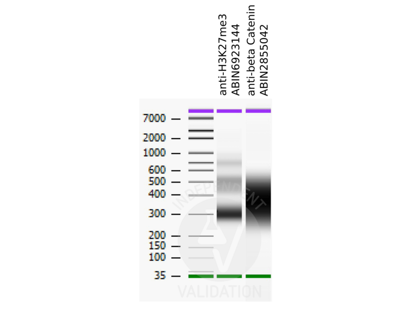
![Bioanalyzer profiles comparing fragment size distributions between reads obtained from CUT&RUN using an anti-H3K27me3 CUT&RUN Positive Control antibody ABIN6923144 and anti-beta Catenin ABIN2855042 after library preparation.]() Bioanalyzer profiles comparing fragment size distributions between reads obtained from CUT&RUN using an anti-H3K27me3 CUT&RUN Positive Control antibody ABIN6923144 and anti-beta Catenin ABIN2855042 after library preparation.
Bioanalyzer profiles comparing fragment size distributions between reads obtained from CUT&RUN using an anti-H3K27me3 CUT&RUN Positive Control antibody ABIN6923144 and anti-beta Catenin ABIN2855042 after library preparation.
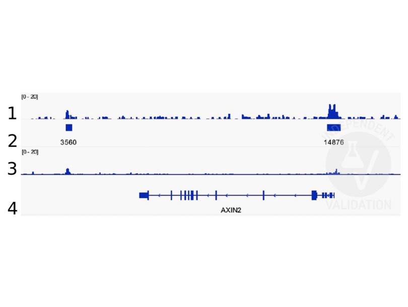
![Alignment tracks from CUT&RUN targeting beta Catenin in HEK293T cells. 1. Alignment track for CUT&RUN reads obtained using anti-beta Catenin antibody ABIN2855042 in HEK293T cells. 2. Peaks called by SEACR from CUT&RUN data using anti-beta Catenin antibody ABIN2855042. 3. Peak track from ChIP-seq targeting beta Catenin in HEK293T cells (Doumpas et al., 2019). 4. Refseq track showing the AXIN2 gene on (chr17:65528563-65561648).]() Alignment tracks from CUT&RUN targeting beta Catenin in HEK293T cells. 1. Alignment track for CUT&RUN reads obtained using anti-beta Catenin antibody ABIN2855042 in HEK293T cells. 2. Peaks called by SEACR from CUT&RUN data using anti-beta Catenin antibody ABIN2855042. 3. Peak track from ChIP-seq targeting beta Catenin in HEK293T cells (Doumpas et al., 2019). 4. Refseq track showing the AXIN2 gene on (chr17:65528563-65561648).
Full Methods
Alignment tracks from CUT&RUN targeting beta Catenin in HEK293T cells. 1. Alignment track for CUT&RUN reads obtained using anti-beta Catenin antibody ABIN2855042 in HEK293T cells. 2. Peaks called by SEACR from CUT&RUN data using anti-beta Catenin antibody ABIN2855042. 3. Peak track from ChIP-seq targeting beta Catenin in HEK293T cells (Doumpas et al., 2019). 4. Refseq track showing the AXIN2 gene on (chr17:65528563-65561648).
Full Methods -
- Format
- Liquid
- Concentration
- 0.07 mg/mL
- Buffer
- 1XPBS ( pH 7), 1 % BSA, 20 % Glycerol, 0.025 % ProClin 300
- Preservative
- ProClin
- Precaution of Use
- This product contains ProClin: a POISONOUS AND HAZARDOUS SUBSTANCE which should be handled by trained staff only.
- Storage
- 4 °C,-20 °C
- Storage Comment
- Store as concentrated solution. Centrifuge briefly prior to opening vial. For short-term storage (1-2 weeks), store at 4°C. For long-term storage, aliquot and store at -20°C or below. Avoid multiple freeze-thaw cycles.
-
-
: "Acute Intoxication With Alcohol Reduces Trauma-Induced Proinflammatory Response and Barrier Breakdown in the Lung via the Wnt/β-Catenin Signaling Pathway." in: Frontiers in immunology, Vol. 13, pp. 866925, (2022) (PubMed).
: "A new cut&run low volume-urea (LoV-U) protocol optimized for transcriptional co-factors uncovers Wnt/b-catenin tissue-specific genomic targets." in: Development (Cambridge, England), (2022) (PubMed).
: "Identification of candidate lncRNAs and circRNAs regulating WNT3/β-catenin signaling in essential hypertension." in: Aging, Vol. 12, Issue 9, pp. 8261-8288, (2020) (PubMed).
: "A novel c-Kit/phospho-prohibitin axis enhances ovarian cancer stemness and chemoresistance via Notch3-PBX1 and β-catenin-ABCG2 signaling." in: Journal of biomedical science, Vol. 27, Issue 1, pp. 42, (2020) (PubMed).
: "Small G protein signalling modulator 2 (SGSM2) is involved in oestrogen receptor-positive breast cancer metastasis through enhancement of migratory cell adhesion via interaction with E-cadherin." in: Cell adhesion & migration, Vol. 13, Issue 1, pp. 120-137, (2019) (PubMed).
: "Targeting the Wnt/β-catenin pathway in human osteosarcoma cells." in: Oncotarget, Vol. 9, Issue 95, pp. 36780-36792, (2018) (PubMed).
: "ST6GALNAC1 plays important roles in enhancing cancer stem phenotypes of colorectal cancer via the Akt pathway." in: Oncotarget, Vol. 8, Issue 68, pp. 112550-112564, (2017) (PubMed).
: "Control of the negative IRES trans-acting factor KHSRP by ubiquitination." in: Nucleic acids research, Vol. 45, Issue 1, pp. 271-287, (2017) (PubMed).
-
: "Acute Intoxication With Alcohol Reduces Trauma-Induced Proinflammatory Response and Barrier Breakdown in the Lung via the Wnt/β-Catenin Signaling Pathway." in: Frontiers in immunology, Vol. 13, pp. 866925, (2022) (PubMed).
-
- Target
- CTNNB1 (Catenin (Cadherin-Associated Protein), beta 1, 88kDa (CTNNB1))
- Alternative Name
- catenin beta 1 (CTNNB1 Products)
- Background
-
Beta-catenin is an adherens junction protein. Adherens junctions (AJs, also called the zonula adherens) are critical for the establishment and maintenance of epithelial layers, such as those lining organ surfaces. AJs mediate adhesion between cells, communicate a signal that neighboring cells are present, and anchor the actin cytoskeleton. In serving these roles, AJs regulate normal cell growth and behavior. At several stages of embryogenesis, wound healing, and tumor cell metastasis, cells form and leave epithelia. This process, which involves the disruption and reestablishment of epithelial cell-cell contacts, may be regulated by the disassembly and assembly of AJs. AJs may also function in the transmission of the 'contact inhibition' signal, which instructs cells to stop dividing once an epithelial sheet is complete.[supplied by OMIM]
Cellular Localization: Cytoplasm , Nucleus , cytoskeleton - Molecular Weight
- 85 kDa
- Gene ID
- 1499
- UniProt
- P35222
- Pathways
- WNT Signaling, Intracellular Steroid Hormone Receptor Signaling Pathway, Peptide Hormone Metabolism, Regulation of Muscle Cell Differentiation, Cell-Cell Junction Organization, Tube Formation, Maintenance of Protein Location, Signaling Events mediated by VEGFR1 and VEGFR2
-


