Phone:
+1 877 302 8632
Fax:
+1 888 205 9894 (Toll-free)
E-Mail:
orders@antibodies-online.com

IHC Antibodies

antibodies-online offers you currently more than 200,000 IHC antibodies for a wide variety of targets.

Antibodies for IHC

Overview of Immunohistochemistry

Immunohistochemistry (IHC) refers to the process of detecting antigens in cells of a tissue section by exploiting the principle of antibodies binding specifically to antigens in biological tissues. Its widely used in the diagnosis of abnormal cells such as those found in cancerous tumors and in basic research to understand the distribution and localization of biomarkers and differentially expressed proteins in different parts of a biological tissue. Visualising an antibody-antigen interaction can be accomplished in a number of ways. In the most common instance, an antibody is conjugated to an enzyme, such as peroxidase, that can catalyse a colour-producing reaction or tagged to a fluorophore.

anti-Collagen Type I antibody ABIN5596819

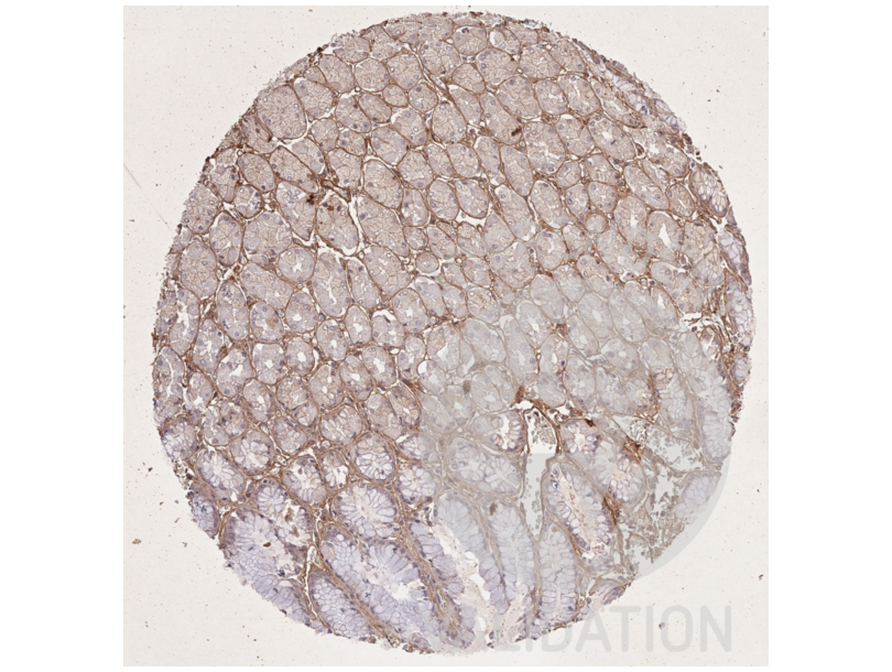
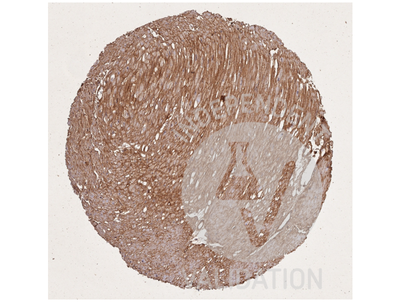
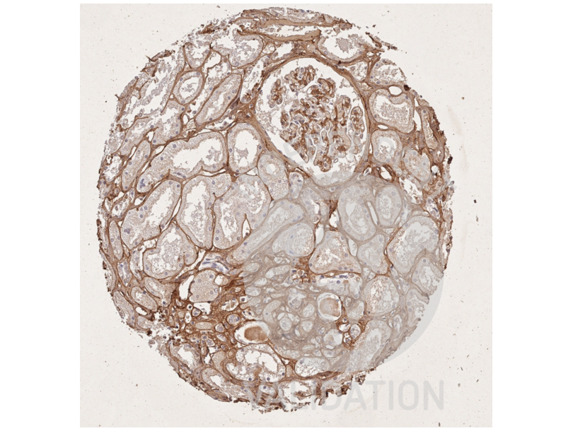
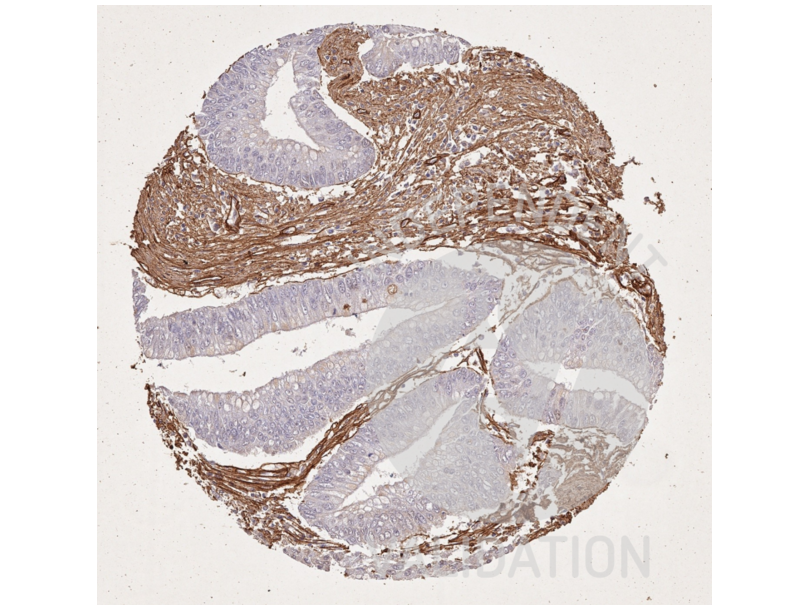
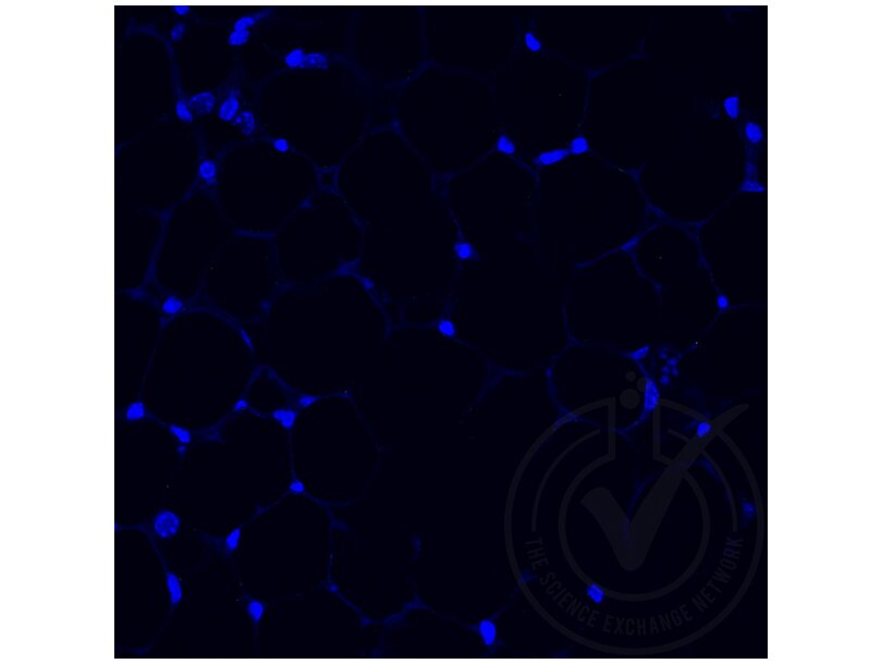
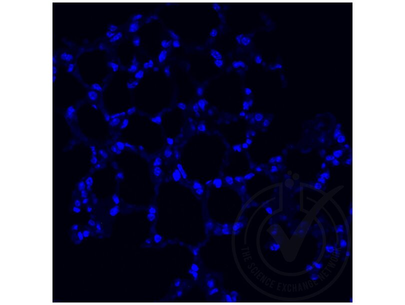
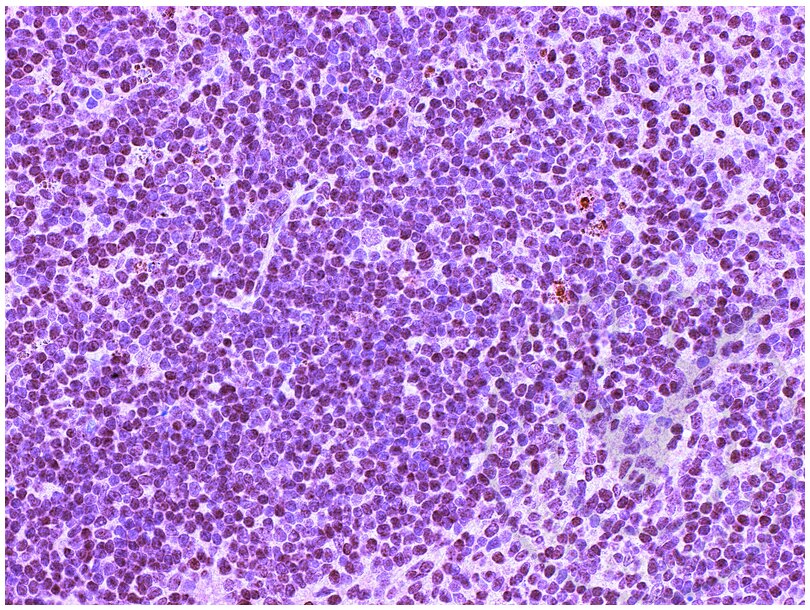
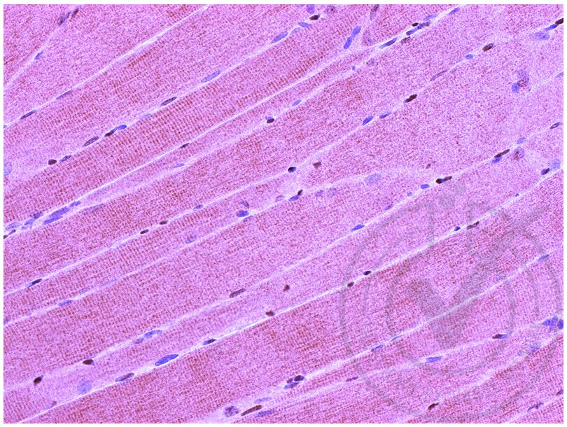
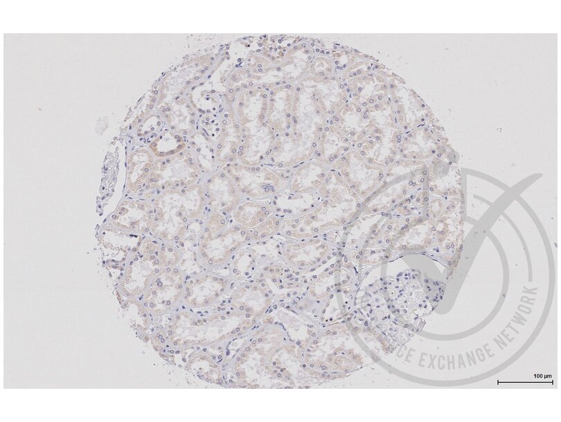
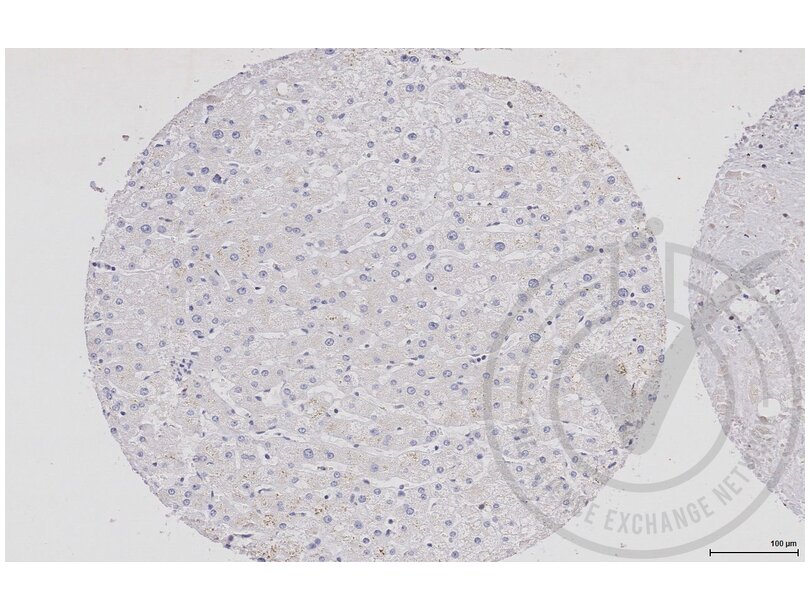
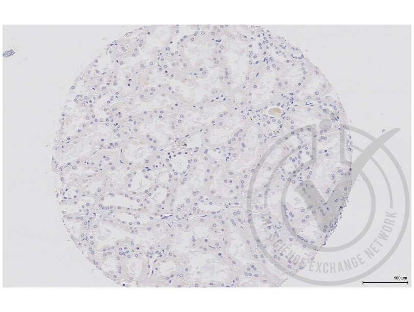
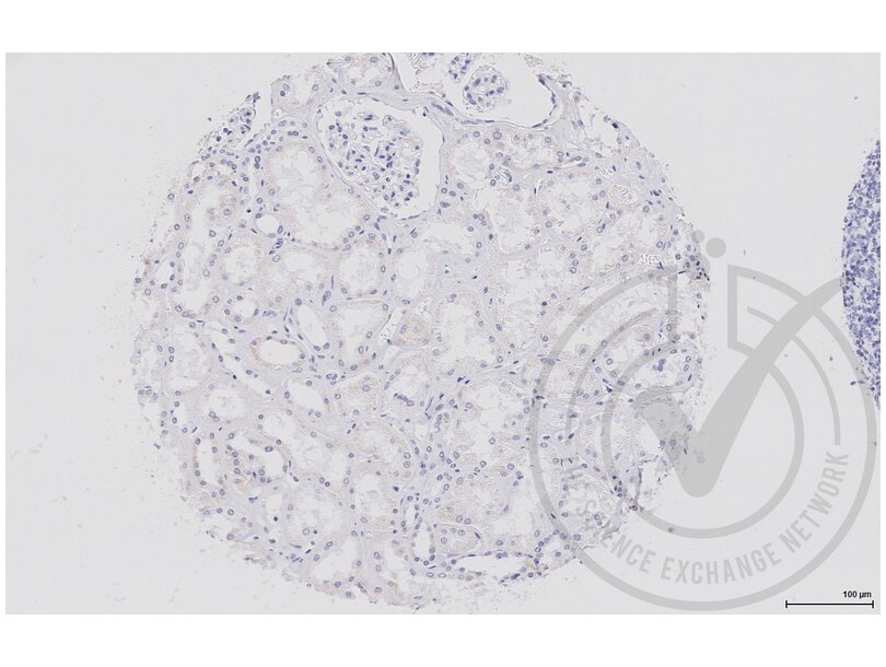
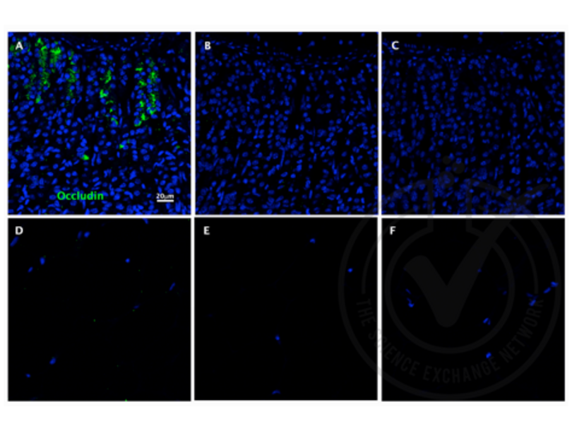
ABIN5596819 : anti-Collagen Type I antibody validated for IHC

Learn more about Immunohistochemistry (IHC)

Tissue Preparation

The tissue sample preparation is of crucial importance, because inadequate handling may disrupt tissue structure, cause reduced antibody binding affinity or even prevent binding altogether. The goal is to preserve the tissue in close to life-like conditions while concomitantly preventing autolysis and degradation due to bacterial or fungal growth. To this end, fixatives are used, such as paraformaldehyde-lysin-periodate (PLP) or formalin. The most common fixative is 4%-10% (in some cases up to 40%) formaldehyde in 0.1M phosphate buffer (in order to stabilize the pH).

For tissue that does not tolerate formaldehyde incubation, an often used alternative is that samples are snap-frozen in liquid nitrogen, and then cut into sections. Tissues incubated with fixatives are embedded in a matrix, e.g. paraffin, or, after they have been dehydrated, in alcohol (isopropanol or ethanol), and then cut into sections. For sensitive tissues the application of a vibratome microtome may be advisable, due to the lower physical stress it produces on the tissues.

Antigen Retrieval

Despite its superior preservation quality, in terms of morphology, formalin-fixed paraffin-embedded samples can lose some or all immunoreactivity due to structural changes of the targeted antigen. The demonstration of antigens, and hence the immunoreactivity, can be improved using various techniques. Mainly, there are two distinct approaches, i.e. heat treatment, the so-called "heat induced epitope retrieval" (HIER), and the "proteolytic induced epitope retrieval" (PIER). In order to apply these techniques tissue-sections have to be deparaffinized and rehydrated first.

HIER works by applying, what is often referred to as retrieval solution. This preheated buffer has varying composition and pH, often consisting of Tris-HCl or citrate. The emerged samples are exposed to heat for varying times (usually between 10-60 minutes) and then slowly cooled. The heating itself may be carried out using an autoclave, waterbath, pressure-cooker or similar appliance.

PIER works by applying digestion enzymes, such as proteinase K, trypsin, pepsin and various other proteinases. Optimal incubation time and concentrations have to be tested. In the final run sample specificities should be optimized. Occasionally it may be required to combine HIER and PIER in order to achieve antigen "unmasking" following formalin-incubation.

Staining Methodologies

Direct method

In this method a labelled antibody (e.g. with a substrate-chromogen) reacts directly with the antigen in the tissue. The advantage is that only one antibody is needed, hence the application is fast and produces little nonspecific binding. On the other hand, since only one antibody binds one epitope, the signal intensity is low. For applications with small amounts of antigen the signal may be too weak.

Indirect method (two-step)

In order to upscale the low signal intensity of the direct method, the indirect method has been developed. The primary antibody binds to the antigen, followed by a second (labelled) antibody binding the primary. The signal is amplified due to the binding of multiple secondary antibodies to a single primary antibody. Another advantage is that only one labelled antibody needs to be employed for different targets, which may reduce costs. One disadvantage is that nonspecific binding occurs more frequently than with the direct method.

Three-step method

In order to further amplify the signal an additional antibody may be employed that binds to the secondary antibody. This method leads to a third layer of antibodies all of which are labelled. This application is useful for staining of antigens with a limited number of epitopes.

PAP method

Today, the peroxidase anti-peroxidase method is rarely used, but has been popular in pathology laboratories previously. It is an indirect method which depends on a third layer of antibodies, bound to peroxidase, that binds an unconjugated second layer antibody. The peroxidase is not chemically conjugated to the IgG but immunologically bound. That means that the third layer antibody is specific for peroxidase. This leads to a much higher activity of the peroxidase, in turn increasing the assay sensitivity by two to three orders of a magnitude.

(Strept)Avidin-Biotin Complex (ABC) methods

Nowadays, one of the most commonly used method for staining is based on the high affinity that avidin (chicken egg) and streptavidin (Streptomyces avidinii) have for the glycoprotein biotin. The basic principle is that avidin (or streptavidin) reacts with a biotinylated secondary antibody, followed by a horseradish peroxidase reaction.

In some cases catalyzed signal amplification (CSA) is employed. An amplification reagent leads to the aggregation of a large number of biotins that subsequently react with streptavidin-labelled peroxidase. One issue with avidin is its large electrostatic binding, due to its molecular charge. Streptavidin does not have the same propensity, thus reducing the background signal.

Polymeric methods

In short, polymeric methods employ large antibody-bound polymers with the ability to bind a larger number of molecules, typically an average of ten antibodies and approximately 70 enzyme molecules. This setup allows for outstanding amplification of the signal, hence high sensitivity, reduced nonspecific binding, and therefore low background signal. It also allows for staining of two different antigens at the same time.

222,491 results :

RFP Reactivity: Discosoma WB, ELISA, IF, IP, IHC, FACS, IHC (fro), IHC (p) Host: Rabbit Polyclonal unconjugated
Pubmed 356 references Independent Validation 1 validation
Catalog No. ABIN129578
Catalog No. ABIN724340
CDH1 Reactivity: Human, Mouse, Rat WB, FACS, ELISA, IHC (p), ICC, IHC (fro), IF (p), IF (cc) Host: Rabbit Polyclonal unconjugated
Pubmed 14 references Independent Validation 1 validation
Catalog No. ABIN1387847
TNF alpha Reactivity: Human, Mouse, Rat, Rabbit, Horse WB, ELISA, IHC (p), IF (p), IHC (fro) Host: Rabbit Polyclonal unconjugated
Pubmed 21 references Independent Validation 1 validation
Catalog No. ABIN677318
CASP8 Reactivity: Human, Mouse, Rat, Monkey, Pig, Sheep WB, ELISA, IHC (p), FACS, IF (cc), IF (p), IHC (fro) Host: Rabbit Polyclonal unconjugated
Pubmed 15 references Independent Validation 1 validation
Catalog No. ABIN724205
CD63 Reactivity: Human, Mouse, Rat, Monkey, Cow, Dog, Chicken, Pig, Rabbit, Donkey, Sheep, Goat, Guinea Pig, Avian, Hamster, Cat, Horse FACS, WB, IHC, IF Host: Goat Polyclonal unconjugated
Pubmed 10 references Independent Validation 3 validations
Catalog No. ABIN1440014
KO Validated SOX2 Reactivity: Human WB, IF, FACS, IHC (p), ICC, IP, IHC (fro), CUT&RUN Host: Rabbit Polyclonal unconjugated
Pubmed 11 references Independent Validation 1 validation
Catalog No. ABIN2855074
IBA1 Reactivity: Human WB, IHC, IHC (p), ICC, IF, FACS, IHC (fro), IHC (ff) Host: Rabbit Polyclonal unconjugated
Pubmed 13 references Independent Validation 1 validation
Catalog No. ABIN2857032
  • Type Primary
    • Primary
  • Target
    • Histone 3 (H3)
    • p53 (TP53)
    • AKT1
    • EGFR
    • CD45 (PTPRC)
    • Albumin (ALB)
    • NF-kB p65 (NFkBP65)
    • Bcl-2 (BCL2)
    • TNF alpha
    • Estrogen Receptor alpha (ESR1)
    • GFAP
    • CD4
    • HSP70
    • Vimentin (VIM)
    • EpCAM (EPCAM)
    • C-JUN (JUN)
    • MUC1
    • Cytokeratin 18 (KRT18)
    • c-MYC (MYC)
    • ErbB2/Her2
    • KRT8
    • CD8 alpha (CD8A)
    • Retinoblastoma 1 (RB1)
    • tau
    • BAD
    • Chromogranin A (CHGA)
    • HSP27 (HSPB1)
    • HSPD1
    • Cytokeratin 19 (KRT19)
    • CD31 (PECAM1)
    • SMAD3
    • SNCA
    • CD34
    • Caspase 3 (CASP3)
    • RPS6KB1
    • CD44
    • alpha Fetoprotein (AFP)
    • Insulin (INS)
    • MEK1 (MAP2K1)
    • RAF1
    • KIT
    • MLANA
    • SOD1
    • APP
    • Cytokeratin 7 (KRT7)
    • p21 (CDKN1A)
    • Interferon gamma (IFNG)
    • FAK (PTK2)
    • ATF2
    • ABL1
    Please use search to find more...
  • Application Immunohistochemistry (IHC), Immunohistochemistry (Paraffin-embedded Sections) (IHC (p)), Immunohistochemistry (Frozen Sections) (IHC (fro)), Immunohistochemistry (Formalin-fixed Sections) (IHC (f)), Immunohistochemistry (Formalin-fixed Paraffin-embedded Sections) (IHC (fp)), Immunohistochemistry (Zinc-fixed Sections) (IHC (zinc)), Immunohistochemistry (Paraformaldehyde) (IHC (pfa)), Immunohistochemistry (Free Floating) (IHC (ff)), Immunohistochemistry (Whole Mount) (IHC (wm)), Immunohistochemistry (Resin Sections) (IHC (rs)), Immunohistochemistry (Fixed) (IHC (fx)), Immunohistochemistry (Acetone-fixed) (IHC (af))
    • Immunohistochemistry (Acetone-fixed) (IHC (af))
    • Immunohistochemistry (Fixed) (IHC (fx))
    • Immunohistochemistry (Resin Sections) (IHC (rs))
    • Immunohistochemistry (Whole Mount) (IHC (wm))
    • Immunohistochemistry (Free Floating) (IHC (ff))
    • Immunohistochemistry (Paraformaldehyde) (IHC (pfa))
    • Immunohistochemistry (Zinc-fixed Sections) (IHC (zinc))
    • Immunohistochemistry (Formalin-fixed Paraffin-embedded Sections) (IHC (fp))
    • Immunohistochemistry (Formalin-fixed Sections) (IHC (f))
    • Immunohistochemistry (Frozen Sections) (IHC (fro))
    • Immunohistochemistry (Paraffin-embedded Sections) (IHC (p))
    • Immunohistochemistry (IHC)
    • Western Blotting (WB)
    • ELISA
    • Immunofluorescence (Paraffin-embedded Sections) (IF (p))
    • Immunofluorescence (Cultured Cells) (IF (cc))
    • Flow Cytometry (FACS)
    • Immunofluorescence (IF)
    • Immunoprecipitation (IP)
    • Immunocytochemistry (ICC)
    • Enzyme Immunoassay (EIA)
    • Immunofluorescence (fixed cells) (IF/ICC)
    • Immunochromatography (IC)
    • Dot Blot (DB)
    • Radioimmunoassay (RIA)
    • Functional Studies (Func)
    • Neutralization (Neut)
    • Coating (Coat)
    • Immunostaining (ISt)
    • Staining Methods (StM)
    • Chromatin Immunoprecipitation (ChIP)
    • Immunodiffusion (ID)
    • Immunoassay (IA)
    • Proximity Ligation Assay (PLA)
    • Lateral Flow (LF)
    • Confocal Microscopy (CM)
    • Fluorescence Microscopy (FM)
    • Immunoelectrophoresis (IEP)
    • Indirect Immunofluorescence Assay (IFA)
    • Antibody Array (AA)
    • RNA Interference (RNAi)
    • ChIP DNA-Sequencing (ChIP-seq)
    • Protein Assay (PrA)
    • ELISA (Capture)
    • Cellular Assay (CA)
    • Blocking Peptide (BP)
    • Blocking Reagent (BR)
    • Cytotoxicity Test (CyTox)
    • Live Cell Imaging (LCI)
    • ELISpot
    • Immunoelectron Microscopy (IEM)
    • FLISA
    • Luminex Assay (LMNX)
    • ELISA (Detection)
    • Lateral Flow Chromatographic Immunoassay (LFCIA)
    • Gel Shift (GS)
    • Chemiluminescence Immunoassay (CLIA)
  • Reactivity
    • Human
    • Mouse
    • Rat
    • Cow
    • Dog
    • Monkey
    • Rabbit
    • Pig
    • Horse
    • Guinea Pig
    • Chicken
    • Zebrafish (Danio rerio)
    • Hamster
    • Sheep
    • Bat
    • Xenopus laevis
    • Gorilla
    • Goat
    • Mammalian
    • Cat
    • Saccharomyces cerevisiae
    • Drosophila melanogaster
    • Chimpanzee
    • Frog
    • Fish
    • Non-Human Primate
    • Primate
    • Gibbon
    • Rhesus Monkey
    • Orang-Utan
    • Various Species
    • Virus
    • Plant
    • Cynomolgus
    • Baboon
    • Chemical
    • E. coli
    • Hepatitis C Virus (HCV)
    • Bacteria
    • All Species
    • Aequorea victoria
    • Human Immunodeficiency Virus (HIV)
    • Macaque
    • Avian
    • C. elegans
    • Insect
    • Ferret
    • Human Papillomavirus 16 (HPV-16)
    • General
    • Marmoset
    Please use search to find more...
  • Cited in publications
  • Images available
  • Independent Validation
  • Preservative free only
  • Carrier free only
  • Detection Method
    • Fluorometric
  • Host
    • Rabbit
    • Mouse
    • Goat
    • Rat
    • Sheep
    • Guinea Pig
    • Chicken
    • Hamster
    • Armenian Hamster
    • Human
    • Chicken eggs
    • Horse
    • Humanized
    • Pig
    • Alpaca
    • Cell culture
    • Chicken egg
    • Golden Syrian Hamster
    • Rabbit/Mouse Cocktail
  • Clonality
    • Polyclonal
    • Monoclonal
    • Chimeric
  • Clone
    • C1
    • C2
    • C3
    • C5
    • C4
    • D4
    • 9E10
    • 5A5
    • 2F11
    • C6
    • 3F8
    • 2C1
    • 2F6
    • 1F6
    • 1A4
    • 3G4
    • RB6-8C5
    • 3B5
    • 1F8
    • 2A4
    • 2D2
    • 4C2
    • 7F8
    • S153-5
    • 1F10
    • 3G8
    • 6G9
    • C11
    • H57-597
    • SV5-P-K
    • 1A1
    • 1D11
    • 1D4
    • 4A3
    • C7
    • Hi52C
    • PAb
    • UCHT1
    • 1G4
    • 2G4
    • AE-1
    • 1B9
    • 1C7
    • 2C4
    • 2D4
    • 3F4
    • 3G1
    • 8C8
    • D1
    • S75-33
    Please use search to find more...
  • Binding Specificity
    • C-Term
    • N-Term
    • Internal Region
    • Center
    • AA 1-30
    • AA 1-100
    • Middle Region
    • AA 101-200
    • AA 51-150
    • AA 201-300
    • Extracellular Domain
    • AA 21-120
    • AA 151-250
    • AA 1-50
    • AA 301-400
    • Cytoplasmic Domain
    • AA 251-350
    • AA 31-130
    • AA 401-500
    • AA 351-450
    • Intracellular
    • AA 1-300
    • AA 501-600
    • AA 1-80
    • AA 451-550
    • AA 121-220
    • AA 1-200
    • AA 131-230
    • Extracellular
    • AA 50-100
    • 3rd Cytoplasmic Domain
    • AA 150-200
    • AA 100-150
    • AA 81-180
    • full length
    • 2nd Extracellular Domain
    • AA 231-330
    • AA 41-140
    • AA 30-110
    • AA 10-90
    • AA 1-250
    • AA 221-320
    • AA 61-160
    • AA 601-700
    • AA 1-280
    • AA 161-260
    • AA 40-120
    • AA 701-800
    • AA 1-260
    • BH3 Domain
    Please use search to find more...
  • Conjugate
    • Un-conjugated
    • Biotin
    • HRP
    • FITC
    • PE
    • APC
    • CF®488A
    • CF®405S
    • CF®568
    • CF®594
    • CF®640R
    • CF®647
    • PerCP
    • Atto 594
    • Alkaline Phosphatase (AP)
    • Atto 488
    • Atto 390
    • Peroxidase (POD)
    • PE-Cy5
    • CF®555
    • PE,Cy5
    • SPRD
    • PE-Cy7
    • Cy5
    • DyLight 488
    • DyLight 550
    • CF®660R
    • Cy7,PE
    • CF405M
    • DyLight 650
    • PE,Cy7
    • APC-Cy7
    • CF®770
    • PE-Cy5.5
    • Cy3
    • PE,Cy5.5
    • CF®543
    • Streptavidin
    • Atto 633
    • TRITC
    • Cy5.5,PE
    • PE-DyLight 594
    • Atto 550
    • CF-Blue
    • CF®680
    • CF®680R
    • GST tag
    • His tag
    • PE,Texas Red (TR)
    • Rhodamine
    • DyLight 405
  • Isotype
    • IgG
    • IgG1
    • Ig Fraction
    • IgG1 kappa
    • IgG2a
    • IgG2b
    • IgG2a kappa
    • IgG2b kappa
    • IgG kappa
    • IgM
    • IgG2
    • IgY
    • IgG3
    • IgM kappa
    • IgG2c kappa
    • IgG3 kappa
    • kappa
    • IgG lambda
    • IgG Mix lambda
    • IgG1 lambda
    • IgG2a lambda
    • IgG2c
    • IgG2b lambda
    • IgG2 kappa
    • IgG1/IgG2b
    • Ig kappa
    • IgA
    • IgG4
    • IgG4 kappa
    • Ig Mix
    • Cocktail
    • IgG1a
    • IgG3 lambda
    • IgA kappa
    • IgE
    • IgG Mix
    • IgG1 lamda
    • IgG2 lambda
    • IgG4 lambda
  • Protein Type
    • Recombinant Antibody
  • Format
    • Liquid
    • Lyophilized
    • Lyophilized or Liquid
    • Frozen
  • Fragment
    • F(ab')2 fragment
    • Fab fragment
    • scFv fragment
    • single-domain Antibody (sdAb)
  • Grade
    • Verified
    • KO Validated
    • Microarray verified
    • GMP Grade
    • In vivo Grade
    • Cell Culture Grade
    • Calibrator Grade
    • ChIP-seq Grade
  • Supplier
    • antibodies-online
    • USBio
    • Abbexa
    • Cloud-Clone
    • Acris
    • Biotium
    • Assay Biotechnology Company, Inc.
    • Covalab
    • NSJ Bioreagents
    • Aviva Systems Biology
    • Cohesion Biosciences
    • Assaypro
    • RayBiotech
    • ProSci
    • FabGennix
    • Boster Bio
    • Signalway
    • Alomone Labs
    • Angio-Proteomie
    • OriGene
    • Wuhan Fine biotech
    • Enogene Biotech
    • Rockland
    • CUSABIO
    • Antibodies Incorporated
    • St John's Laboratory
    • PROGEN
    • Synaptic Systems
    • Randox Lifesciences
    • Elabscience
    • QED Bioscience
    • EnCor Biotechnology
    • Kamiya Biomedicals
    • Neuromics Antibodies
    • Abeomics
    • ANTIGENES
    • Active Motif
    • RevMAb Biosciences
    • ProteoGenix
    • Agrisera
    • BioAcademia
    • Aeonian Biotech
    • Transgenic
    • Ansh Labs
    • ImmunoBioscience
    • Biohit
    • Aldevron
    • Biospes
    • AntiProt
    • Hytest
    Please use search to find more...
You are here:
Support