EGFR antibody (C-Term)

 (3 references)
 (1 validation)
Quick Overview for EGFR antibody (C-Term) (ABIN98862)
Key Features
- High quality EGFR Primary Antibody for the detection of EGFR.
- Reliable product with high standard validation data
Target
See all EGFR AntibodiesReactivity
Host
Clonality
Conjugate
Application
-
-
Binding Specificity
- C-Term
-
Supplier Product No.
- 100-401-149
-
Supplier
- Rockland
-
Purpose
- EGFR Antibody
-
Cross-Reactivity (Details)
- This antiserum is directed against human epidermal growth factor receptor (EGFR) and is useful in determining its presence in western blotting and immunoprecipitation experiments. This antibody can detect EGFR from human, mouse and rat sources.
-
Characteristics
- Synonyms: rabbit anti-EGFR Antibody, rabbit anti-epidermal growth factor receptor antibody, Receptor tyrosine-protein kinase erbB-1 antibody, c-erbB-1 antibody
-
Purification
- Antiserum
-
Sterility
- Sterile filtered
-
Immunogen
-
Immunogen: This whole rabbit serum was prepared by repeated immunizations with a peptide synthesized using conventional technology. The sequence of the epitope maps to a region near the carboxy terminus which is identical in human, mouse and rat EGFR.
Immunogen Type: Conjugated Peptide
-
Product Specific Information
-
- What can the EGFR Antibody ABIN98862 be used for?
- This polyclonal EGFR Antibody detects EGFR. The EGFR Antibody has been validated for various applications and can be used for the detection of EGFR and derivatives by Western Blotting, Immunohistochemistry, ELISA, Immunoprecipitation.
- What validation data is available for this EGFR Antibody?
- The primary antibody is referenced in 3 publications and is characterized by a proven, very high reliability. It has currently 2 product images that show its performance in a variety of applications. The product is currently available in 250 μL quantity. EGFR Antibody for the detection of EGFR and derivatives.
- What is the function of EGFR?
- The protein encoded by this gene is a transmembrane glycoprotein that is a member of the protein kinase superfamily. This protein is a receptor for members of the epidermal growth factor family. EGFR is a cell surface protein that binds to epidermal growth factor. Binding of the protein to a ligand induces receptor dimerization and tyrosine autophosphorylation and leads to cell proliferation. Mutations in this gene are associated with lung cancer. Multiple alternatively spliced transcript variants that encode different protein isoforms have been found for this gene. [provided by RefSeq, Jul 2010].
-
-
anti-Epidermal Growth Factor Receptor (EGFR) (AA 693-893) antibody
EGFR Reactivity: Human WB, IHC, ELISA, FACS Host: Mouse Monoclonal 7A6F12 unconjugated
anti-Epidermal Growth Factor Receptor (EGFR) (AA 888-1210) antibodyEGFR Reactivity: Human WB, IHC Host: Mouse Monoclonal C4 unconjugated
anti-Epidermal Growth Factor Receptor (EGFR) (pSer1071) antibodyEGFR Reactivity: Human, Mouse, Rat WB, IHC, ELISA, IF, ICC Host: Rabbit Polyclonal unconjugated
anti-Epidermal Growth Factor Receptor (EGFR) (pSer1026) antibodyEGFR Reactivity: Human, Mouse, Rat WB, IHC, ELISA, IF, ICC Host: Rabbit Polyclonal unconjugated
anti-Epidermal Growth Factor Receptor (EGFR) (pSer695) antibodyEGFR Reactivity: Human, Mouse, Rat WB, IHC, ELISA, IF, ICC Host: Rabbit Polyclonal unconjugated
anti-Epidermal Growth Factor Receptor (EGFR) (C-Term) antibodyEGFR Reactivity: Human, Mouse, Rat, Monkey, Pig, Cow, Chicken, Dog, Horse, Rabbit, Guinea Pig, Avian, Cat, Donkey, Goat, Hamster, Sheep WB, IHC (p), IHC (fro) Host: Goat Polyclonal unconjugated
anti-Epidermal Growth Factor Receptor (EGFR) (AA 693-893) antibodyEGFR Reactivity: Human WB, ELISA, FACS Host: Mouse Monoclonal 5E10D3 unconjugated
anti-Epidermal Growth Factor Receptor (EGFR) antibodyKD Validated EGFR Reactivity: Human WB, FACS, IF (p) Host: Rabbit Monoclonal 24GB4395 unconjugated Recombinant Antibody
anti-Epidermal Growth Factor Receptor (EGFR) (AA 1163-1191), (C-Term) antibodyEGFR Reactivity: Human WB, IHC (p) Host: Mouse Monoclonal 688CT33-1-3 unconjugated
anti-Epidermal Growth Factor Receptor (EGFR) (AA 888-1210) antibodyEGFR Reactivity: Human WB, IHC, IP, ICC Host: Rabbit Polyclonal unconjugated
-
-
Application Notes
-
Immunohistochemistry Dilution: 2.5 μg/mL
Application Note: Anti-EGFR antibody has been tested by and is specifically designed for ELISA, immunoblotting, immunoprecipitation, and immunohistochemistry. Reactivity in other assays is likely, but has not been determined. Recognition of EGFR is independent of the phosphorylation status at tyrosine 1173. A431 cells, keratinocytes in normal epidermis, or placenta are typically used as positive control sources. The antigen is typically localized in the cell membrane. For western blotting, good results are also achieved on PVDF membranes blocked with 5 % lowfat milk diluted in TTBS for 1 hour at room temperature. Also, dilute the primary antibody and secondary in 5 % lowfat milk in TTBS. Anti-EGFR can be diluted up to 1:10,000 for immunoblot depending on the cell line and the amount of EGFR in a particular lysate. For immunoprecipitation, use approximately 10 μL of the antibody. The immunoprecipitation mix should contain the antibody, 25 μL of Protein A-agarose beads and 1.0 mL of lysate (lysate contains approximately 1.0 mg of total protein). This mixture should be rotated overnight at 4 °C and then washed 3 times with lysis buffer (used to prepare the lysate). The resulting bead complex is dissolved in 20-30 μL of 3X SDS-PAGE sample buffer and approximately 15 μL is loaded per lane on an 8 % polyacrylamide gel.
Western Blot Dilution: 1:1,000 - 1:10,000
Immunoprecipitation Dilution: 10 μL
ELISA Dilution: 1:10,000 - 1:50,000
Other: User Optimized
-
Restrictions
- For Research Use only
-
-
- by
- ADS Biosystems Inc
- No.
- #029817
- Date
- 09/18/2014
- Antigen
- Lot Number
- 19538
- Method validated
- Western Blotting
- Positive Control
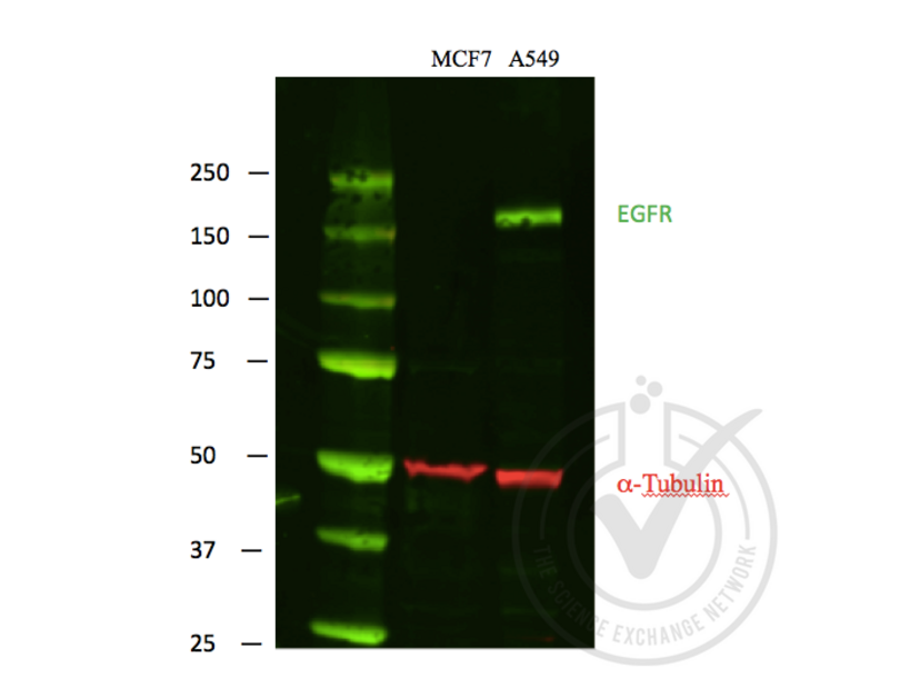
- A549 cells
- Negative Control
- MCF-7 cells
- Notes
- A strong specific band was observed in the positive control at the expected size (~175 kDa) that is not observed in the negative control.
- Primary Antibody
- Antigen: Epidermal Growth Factor Receptor (EGFR)
- Catalog number: ABIN98862
- Lot number: 19538
- Dilution: 1:1,000
- Secondary Antibody
- Antibody: IRDye 680LT Goat Anti-Rabbit
- Lot number: C30725-01
- Dilution: 1:10,000
- Full Protocol
- Lysates were mixed with NuPAGE® LDS Sample Buffer (Life Technologies NP0007) and NuPAGE® Sample Reducing Agent (Life Technologies NP0004) and denatured for 5 minutes at 90ºC.
- 40 μg of each lysate was electrophoresed on a Bolt 4-12% Bis-Tris Gel (Life Technologies BG04120BOX) and run in Bolt MOPS SDS Running Buffer (Life Technologies B0001) at 160 volts for 1 hour.
- Odyssey Western Protein Standard (LI-COR #928-40000) was run as a molecular weight standard.
- PVDF membrane was activated with methanol.
- Protein samples were transferred to activated PVDF membrane in a wet Bolt Transfer Apparatus (Life Technologies B1000) at room temperature for 1 hour at 20 volts (started at 230mA, ended at 110mA).
- The membrane was blocked in x LI-COR Odyssey WB block solution for 1 hour at room temperature.
- The membrane was incubated with the primary antibody diluted 1:1000 in x LI-COR Odyssey WB block solution incubated 2 hours at room temperature.
- The membrane was washed 4 x 5 minutes in 1 x PBS-T (PBS solution with 0.1% Tween 20).
- The membrane was incubated with IRDye® 800CW Goat anti-Mouse Secondary Antibody (Red) and IRDye 680LT Goat Anti-Rabbit Secondary Antibody (Green) from LI-COR (#827-11081, Lot #C30725-01), both 1:10,000 dilutions. Incubation was performed at room temperature for 45 minutes.
- The membrane was washed 4 x 5 minutes in 1 x PBS-T (PBS solution with 0.1% Tween 20).
- Proteins were detected using Odyssey machine scanning with green channel for loading control and red channel for potential LPL band.
- Experimental Notes
- - No experimental challenges noted.
Validation #029817 (Western Blotting)
 Validation Images
![Figure 1: Scanned image of EGFR (Green) and loading control alpha-tubulin (Red) Western blot using LI-COR Odyssey Infrared Technology. First lane, protein molecular weight markers. Second lane, MCF-7 negative control lysate. Third lane, A549 positive control lysate.]() Figure 1: Scanned image of EGFR (Green) and loading control alpha-tubulin (Red) Western blot using LI-COR Odyssey Infrared Technology. First lane, protein molecular weight markers. Second lane, MCF-7 negative control lysate. Third lane, A549 positive control lysate.
Full Methods
Figure 1: Scanned image of EGFR (Green) and loading control alpha-tubulin (Red) Western blot using LI-COR Odyssey Infrared Technology. First lane, protein molecular weight markers. Second lane, MCF-7 negative control lysate. Third lane, A549 positive control lysate.
Full Methods -
-
Format
- Liquid
-
Concentration
- 85 mg/mL
-
Buffer
-
Buffer: None
Stabilizer: None
Preservative: 0.01 % (w/v) Sodium Azide -
Preservative
- Sodium azide
-
Precaution of Use
- This product contains Sodium azide: a POISONOUS AND HAZARDOUS SUBSTANCE which should be handled by trained staff only.
-
Storage
- 4 °C,-20 °C
-
Storage Comment
- Store vial at -20° C prior to opening. Aliquot contents and freeze at -20° C or below for extended storage. Avoid cycles of freezing and thawing. Centrifuge product if not completely clear after standing at room temperature. This product is stable for several weeks at 4° C as an undiluted liquid. Dilute only prior to immediate use.
-
Expiry Date
- 12 months
-
-
-
: "Sensitivity of human granulosa cell tumor cells to epidermal growth factor receptor inhibition." in: Journal of molecular endocrinology, Vol. 52, Issue 2, pp. 223-34, (2014) (PubMed).
: "Evaluation of the cytotoxic effects of ophthalmic solutions containing benzalkonium chloride on corneal epithelium using an organotypic 3-D model." in: BMC ophthalmology, Vol. 9, pp. 5, (2009) (PubMed).
: "Downregulation of EGF receptor signaling in pancreatic islets causes diabetes due to impaired postnatal beta-cell growth." in: Diabetes, Vol. 55, Issue 12, pp. 3299-308, (2007) (PubMed).
-
-
- EGFR (Epidermal Growth Factor Receptor (EGFR))
-
Alternative Name
- EGFR
-
Background
- Background: EGFR is a transmembrane glycoprotein that is a member of a family of protein tyrosine kinases crucial to maintaining a normal balance in cell growth and development. Growth factor receptors are involved not only in promoting the proliferation of normal cells but also in the aberrant growth of many types of human tumors. For example, the epidermal growth factor receptor (EGFR) is mutated and/or over-expressed in many common solid human squamous cell carcinomas including breast, brain, bladder, lung, gastric, head & neck, esophagus, cervix, vulva, ovary, and endometrium. Over-expression of the EGFR gene occurs in carcinomas with and without gene amplification. EGFR and ErbB-2 are particularly important in breast cancer because increased production or activation has been associated with poor prognosis. EGFR belongs to a family of growth factor receptors, which also includes ErbB-2/HER-2/neu, ErbB-3/HER-3/neu and ErbB-4/HER-4/neu. EGFR can heterodimerize with each of the members of this family.
-
Gene ID
- 1956, 29725609
-
UniProt
- P00533
-
Pathways
- NF-kappaB Signaling, RTK Signaling, Fc-epsilon Receptor Signaling Pathway, EGFR Signaling Pathway, Neurotrophin Signaling Pathway, Stem Cell Maintenance, Hepatitis C, Positive Regulation of Response to DNA Damage Stimulus, Interaction of EGFR with phospholipase C-gamma, Thromboxane A2 Receptor Signaling, EGFR Downregulation, S100 Proteins
Target
-