CD200 antibody (Internal Region)
 (1 reference)
 (1 validation)
Quick Overview for CD200 antibody (Internal Region) (ABIN3187966)
Target
See all CD200 AntibodiesReactivity
Host
Clonality
Conjugate
Application
-
-
Binding Specificity
- Internal Region
-
Specificity
- CD200 Polyclonal Antibody detects endogenous levels of CD200 protein.
-
Characteristics
- Rabbit Polyclonal to CD200.
-
Purification
- The antibody was affinity-purified from rabbit antiserum by affinity-chromatography using epitope-specific immunogen.
-
Immunogen
- Synthesized peptide derived from the Internal region of human CD200 (aa 171-220)
-
Isotype
- IgG
-
-
anti-CD200 (CD200) (AA 41-140) antibody
CD200 Reactivity: Human WB, ELISA, IHC (fro), IF (cc), IHC (p), IF (p) Host: Rabbit Polyclonal unconjugated
anti-CD200 (CD200) (AA 31-232) antibodyCD200 Reactivity: Human WB, IHC Host: Rabbit Polyclonal unconjugated
anti-CD200 (CD200) antibody (PE)CD200 Reactivity: Human FACS Host: Mouse Monoclonal OX-104 PE
anti-CD200 (CD200) antibody (FITC)CD200 Reactivity: Human FACS Host: Mouse Monoclonal OX-104 FITC
anti-CD200 (CD200) antibodyCD200 Reactivity: Human FACS, IHC (fro), IHC (p) Host: Mouse Monoclonal OX-104 unconjugated
anti-CD200 (CD200) (AA 56-257) antibodyCD200 Reactivity: Human ELISA Host: Mouse Monoclonal 6E8B11 unconjugated
anti-CD200 (CD200) antibody (APC)CD200 Reactivity: Human FACS Host: Mouse Monoclonal OX-104 APC
anti-CD200 (CD200) (AA 31-232) antibodyCD200 Reactivity: Human FACS, ELISA Host: Rabbit Monoclonal DM156 unconjugated
anti-CD200 (CD200) antibodyCD200 Reactivity: Mouse, Rat WB Host: Rabbit Polyclonal unconjugated
anti-CD200 (CD200) (AA 34-124) antibodyCD200 Reactivity: Human WB, ELISA Host: Mouse Polyclonal unconjugated
-
-
Application Notes
- WB 1:500-1:2000, IHC-P 1:100-1:300, ELISA 1:20000,
-
Restrictions
- For Research Use only
-
-
- by
- David A. Clark, Department of Medicine and Department of Pathology and Molecular Medicine, McMaster University
- No.
- #103457
- Date
- 11/30/2018
- Antigen
- CD200
- Lot Number
- 6839401
- Method validated
- Immunohistochemistry
- Positive Control
Human 7 week elective termination placenta
- Negative Control
KLH antibody ABIN401183
Diluent
- Notes
Passed. The CD200 antibody ABIN318966 specifically recognizes its antigen in human placental tissue.
- Primary Antibody
- ABIN3187966
- Secondary Antibody
- Bond Polymer Refine Detection kit (Leica, DS9800, lot 49232)
- Full Protocol
- Fix human placental tissue in 10% buffered formalin for 24h at RT (22-23°C), process, and embedded in paraffin.
- Cut paraffin blocks with a Leica CM2255 Microtome into 4µm sections.
- Affix sections to positively charged slides and air dry ON at RT.
- Dewax the slides and hydrate on the automated Leica BOND Rx stainer.
- Antigen retrieval on a Leica BOND RX automated stainer using epitope retrieval buffer 2 (Leica, AR9640, lot ER20172).
- Stain slides with primary
- rabbit anti- CD200 antibody (AA 45-95) (Antibodies-online, ABIN761396, lot 980502W),
- rabbit anti-CD200 antibody (AA 170-220) (Antibodies-online, ABIN318966 lot 6839401) (AA 170-220), or
- rabbit anti-KLH antibody (Antibodies-online, ABIN401183, lot 304770)
- diluted 1:200 in Power Vision IHC Super Blocker (Leica, PV6122). The staining protocol incorporates a modified Leica standard protocol IHC-F (which omits the post-primary step) and uses the standard times outlined in the machine protocol.
- Stain sections with Bond Polymer Refine Detection kit (Leica, DS9800, lot 49232) containing peroxidase block, post primary antibody, polymer as well as DAB chromogen and hematoxylin counterstain for times outlined in the standard protocol IHC-F.
- Remove slides from the Leica Bond Rx and then dehydrate in ethanol and clear in xylene.
- Mount slides mounted in Fisher Chemical Permount Mounting Medium (Fisher Scientific, SP15-500, lot 162767).
- Once the slide-coverslip edges are dry, scan the slides using Leica Imagescope (40x) and photograph into jpg files at 400x.
- Experimental Notes
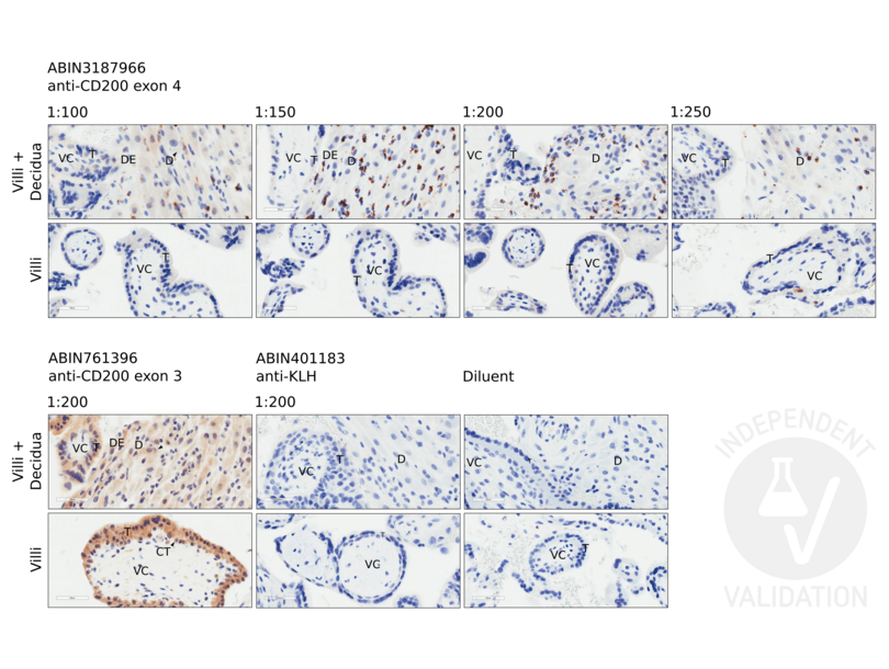
Several dilutions of ABIN318966 were tested and optimal staining was obtained at a dilution as low as 1:200.
The staining pattern of ABIN318966 differs from our result using ABIN761396 which matched RB846 and observations made by others. The result may be explainable by ABIN318966 reacting with variants of CD200 which lack upstream exon 3 coded AA that are eliminated along with exon 2 coded AA by alternative splicing so as to produce the truncated form of CD200 (CD200tr). Removing exon 2 + some exon 3 coded AA may expose epitopes in the downstream exon 3 + exon 4 region of CD200tr that are detected by antibody to the 170-220 AA region of CD200 that are in exon 4 of CD200 variant 1.
ABIN761396 stained villus trophoblasts as described in Clark et al. (2017).
ABIN761396 stained villus cytotrophoblasts and syncytiotrophoblasts but ABIN3187966 did not show convincing staining.
Validation #103457 (Immunohistochemistry)
 Validation Images
![Staining with ABIN318966 at dilutions 1:200, 1:150, 1:200, and 1:250 (two upper rows) using a 7 week elective termination of a missed abortion pregnancy. The upper panels show placental villus and associated decidual tissue, lower panels show staining of villi not associated with decidua. For comparison, tissue samples were stained with ABIN761396, anti-KLH antibody ABIN401183, and diluent alone without any antibody. Images were taken at 400x magnification. VC: placental villus core; T: trophoblast; CT: cytotrophoblast inner layer; ST: syncytiotrophoblast outer layer; DE: decidual epithelium; D: decidua.]() Staining with ABIN318966 at dilutions 1:200, 1:150, 1:200, and 1:250 (two upper rows) using a 7 week elective termination of a missed abortion pregnancy. The upper panels show placental villus and associated decidual tissue, lower panels show staining of villi not associated with decidua. For comparison, tissue samples were stained with ABIN761396, anti-KLH antibody ABIN401183, and diluent alone without any antibody. Images were taken at 400x magnification. VC: placental villus core; T: trophoblast; CT: cytotrophoblast inner layer; ST: syncytiotrophoblast outer layer; DE: decidual epithelium; D: decidua.
Full Methods
Staining with ABIN318966 at dilutions 1:200, 1:150, 1:200, and 1:250 (two upper rows) using a 7 week elective termination of a missed abortion pregnancy. The upper panels show placental villus and associated decidual tissue, lower panels show staining of villi not associated with decidua. For comparison, tissue samples were stained with ABIN761396, anti-KLH antibody ABIN401183, and diluent alone without any antibody. Images were taken at 400x magnification. VC: placental villus core; T: trophoblast; CT: cytotrophoblast inner layer; ST: syncytiotrophoblast outer layer; DE: decidual epithelium; D: decidua.
Full Methods -
-
Format
- Liquid
-
Concentration
- 1 mg/mL
-
Buffer
- Liquid in PBS containing 50 % glycerol, 0.5 % BSA and 0.02 % sodium azide.
-
Preservative
- Sodium azide
-
Precaution of Use
- This product contains Sodium azide: a POISONOUS AND HAZARDOUS SUBSTANCE which should be handled by trained staff only.
-
Handling Advice
- Avoid repeated freeze/thaw cycles.
-
Storage
- -20 °C
-
Storage Comment
- Store at -20°C.
-
-
-
: "CD200S-positive granulated lymphoid cells in endometrium appear to be CD56-positive uterine NK cells." in: Journal of reproductive immunology, Vol. 150, pp. 103477, (2022) (PubMed).
-
: "CD200S-positive granulated lymphoid cells in endometrium appear to be CD56-positive uterine NK cells." in: Journal of reproductive immunology, Vol. 150, pp. 103477, (2022) (PubMed).
-
- CD200
-
Alternative Name
- CD200
-
Molecular Weight
- 31 kDa
-
Gene ID
- 4345
-
UniProt
- P41217
Target
-


