MKRN3 antibody (AA 160-189)
Quick Overview for MKRN3 antibody (AA 160-189) (ABIN651835)
Target
Reactivity
Host
Clonality
Conjugate
Application
Clone
-
-
Binding Specificity
- AA 160-189
-
Purification
- This antibody is purified through a protein A column, followed by peptide affinity purification.
-
Immunogen
- This MKRN3 antibody is generated from rabbits immunized with a KLH conjugated synthetic peptide between 160-189 amino acids from the Central region of human MKRN3.
-
Isotype
- IgG
-
-
-
-
Application Notes
- WB: 1:1000
-
Restrictions
- For Research Use only
-
-
- by
- Department of Medical Biochemistry and Microbiology, Uppsala University
- No.
- #101148
- Date
- 08/10/2017
- Antigen
- MKRN3
- Lot Number
- SA100913BT
- Method validated
- Western Blotting
- Positive Control
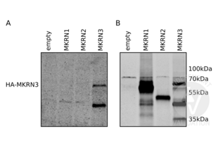
H1299 cells transfected with HA-MKRN3 expression vector; the full-length MKRN3 CDS has been inserted downstream of an HA-tag in plasmid pcDNA3.1
- Negative Control
H1299 cells transfected with HA-MKRN1 or HA-MKRN2 expression plasmid; the full-length MKRN1 or MKRN2 CDS has been inserted downstream of an HA-tag in plasmid pcDNA3
Empty plasmid pcDNA3.1
- Notes
Passed. ABIN2840417 specifically recognizes MKRN3 ectopically expressed in H1299 cells lysates.
- Primary Antibody
- ABIN2840417
- Secondary Antibody
- IRDye 680LT donkey anti-rabbit IgG (H+L) antibody (LI-COR, 925-68023)
- Full Protocol
- Grow H1299 cells (human lung carcinoma cell line) in DMEM medium (ThermoFischer Scientific, 41965-039) supplemented with 10% fetal bovine serum (ThermoFischer Scientific, 10500064) and antibiotics (1X Penicillin-Streptomycin mix, 15140122, ThermoFischer Scientific), at 37°C and 7% CO2 in 12-well plates.
- Transfect H1299 cells with plasmids expressing either HA-MKRN1, HA-MKRN2, HA-MKRN3 or with an empty plasmid pcDNA3.1 (ThermoFischer Scientific, V79020) with max 2µg of plasmid using Turbofect transfection reagent (ThermoFisher Scientific, R0531) following the manufacturer´s instructions.
- Lyse cells in volume RIPA buffer.
- Determine total protein content of the lysates using a Bradford protein assay (Protein Assay Dye Reagent Concentrate, Bio-Rad, 5000006).
- Denature 80µg of total protein for 5min at 95°C in 20µl of 2x Laemmli SDS sample buffer and subsequently separate them on a denaturing Any kD Mini-PROTEAN TGX precast protein gel (Bio-Rad, 4569033) in Mini-PROTEAN Electrophoresis Cells for 1h at 180V.
- Transfer proteins onto Amersham Protran 0.45µm nitrocellulose filter (GE Healthcare Life Sciences) with transfer buffer (25mM Tris, 192mM glycine, 20% methanol) in a Western blotting system for at 4°C for 3h at 120mA.
- Block the membrane with Odyssey Blocking Buffer (LI-COR, 927-50100) for 15min at RT.
- Incubation with primary
- rabbit anti-MKRN3 antibody (antibodies-online, ABIN2840417, lot SA100913BT) diluted 1:1000 in Odyssey Blocking Buffer ON at 4°C or
- mouse anti-HA-tag antibody (BioLegend, 901501) diluted 1:2000 in Odyssey Blocking Buffer ON at 4°C.
- Wash membrane 5x for time with PBS supplemented with 0.1% Tween 20.
- Incubation with secondary
- IRDye 680LT donkey anti-rabbit IgG (H+L) antibody (LI-COR, 925-68023) or
- IRDye 800CW donkey anti-mouse IgG (H+L) antibody (LI-COR, 926-32212)
- diluted 1:10000 in Odyssey Blocking Buffer for 30 min at RT.
- Wash membrane 5x for time with PBS supplemented with 0.1% Tween 20.
- Scan the membrane with LI-COR Odyssey CLX Scanner.
- Experimental Notes
ABIN2840417 is specific towards the expressed HA-MKRN3: it does not recognize HA-MKRN1 (which has 48% identity with MKRN3) and HA-MKRN2 (has 37% identity with MKRN3) ectopically expressed in H1299 cells.
An extraneous band can be detected with the antibody around 40kDa. This band is recognized with both anti HA- and anti MKRN3-antibodies. This suggests either C-terminal degradation product of HA-MKRN3 or that HA-MKRN3 mRNA has been not efficiently translated.
A smear is visible for HA-MKRN3 protein with both antibodies, which may indicate that the protein undergoes self-ubiquitination or forms higher order structures.
At present we have had difficulties to use the antibody to detect endogenous MKRN3 protein. This is probably due to the fact that endogenous MKRN3 protein levels can be very low is some cell lines.
Validation #101148 (Western Blotting)![Successfully validated 'Independent Validation' Badge]()
![Successfully validated 'Independent Validation' Badge]() Validation ImagesFull Methods
Validation ImagesFull Methods -
-
Format
- Liquid
-
Buffer
- Purified polyclonal antibody supplied in PBS with 0.09 % (W/V) sodium azide.
-
Preservative
- Sodium azide
-
Precaution of Use
- This product contains Sodium azide: a POISONOUS AND HAZARDOUS SUBSTANCE which should be handled by trained staff only.
-
Storage
- 4 °C,-20 °C
-
Storage Comment
- Maintain refrigerated at 2-8 °C for up to 6 months. For long term storage store at -20 °C in small aliquots to prevent freeze-thaw cycles.
-
Expiry Date
- 6 months
-
-
-


 (1 validation)


