Nipah Virus
The Nipah virus (NiV) is a zoonotic paramyxovirus associated with recurrent localized outbreaks in South and Southeast Asia. Due to its high case fatality rate (40–75%) and the lack of approved vaccines or specific antiviral therapies, NiV is classified by the World Health Organization (WHO) as a high-priority pathogen within the framework of global research preparedness.
Recent reports of confirmed cases in West Bengal, India, in January 2026 have once again drawn attention to NiV, highlighting the continued importance of surveillance, basic research, and experimental tools.
Nipah Virus Antibodies
Nipah Virus Proteins
Virology and Pathogenesis
NiV belongs to the family Paramyxoviridae, genus Henipavirus. It possesses a single-stranded, negative-sense RNA genome encoding six structural proteins: N (nucleocapsid), P (phosphoprotein), M (matrix protein), F (fusion protein), G (attachment glycoprotein), and L (RNA-dependent RNA polymerase).
The G protein mediates binding to cellular receptors (ephrin-B2 and ephrin-B3), while the F protein catalyzes fusion of the viral envelope with the host cell membrane—key steps in viral entry.
Transmission to humans typically occurs through contact with contaminated food sources (e.g., date palm sap contaminated with fruit bat saliva) or direct exposure to infected animals or individuals. Human-to-human transmission has been documented, particularly in healthcare and caregiving settings.
Epidemiological Situation: Current Outbreaks and Historical Context
In January 2026, Indian authorities confirmed two cases of NiV infection in the state of West Bengal, both involving healthcare workers. A total of 196 contacts were identified; all were asymptomatic and tested negative. Authorities declared the outbreak contained, although several countries in South and Southeast Asia have intensified border controls and health surveillance measures.
Historical outbreak patterns indicate that NiV has repeatedly occurred at a local level in India. In recent years, cases have been reported almost annually in Kerala, with varying case numbers and fatality rates, illustrating the persistent presence and ongoing disease risk in endemic regions.
The virus was first identified during outbreaks in Malaysia and Singapore in 1998–1999. Subsequent cases were repeatedly reported in Bangladesh and India, often associated with spillover events from fruit bats.
Clinical Manifestation and Risk
The incubation period typically ranges from several days to a few weeks. Clinical manifestations range from nonspecific influenza-like symptoms to severe respiratory disease and hemorrhagic encephalitis, which may be fatal.
As no specifically approved treatments or vaccines are currently available, clinical management is limited to supportive care and infection control measures.
Research Needs and Challenges
Despite more than 25 years of knowledge about the pathogen, significant scientific gaps remain, particularly with regard to host–pathogen interactions, immune responses, and therapeutic development. Research in these areas is complex and resource-intensive due to biosafety requirements (BSL-4).
Recent reviews emphasize the need for international harmonization of assays, models, and standardized reagents to ensure reliable results and comparability across laboratories.
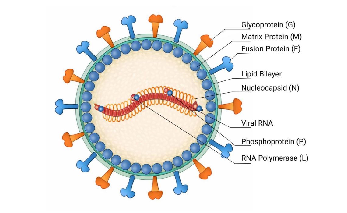
Fig. 1: Schematic, color-coded cross-sectional representation of the Nipah virus with labeled structural components, including lipid bilayer, glycoprotein (G), fusion protein (F), matrix protein (M), nucleocapsid (N), viral RNA, as well as phosphoprotein (P) and RNA polymerase (L).
Experimental Tools: Antibodies and Assays
For non-infectious experimental settings, target-specific antibodies against viral proteins are essential tools:
Antibodies against the G protein (attachment) are used to analyze receptor binding and to characterize surface structures in pseudotyped systems.
Antibodies against the F protein (fusion) enable investigation of membrane fusion mechanisms and are applied in screening assays for entry inhibitors.
Antibodies against the N protein are primarily used for quantitative assessment of viral protein expression in transgenic systems or virological modeling approaches.
Such reagents are validated exclusively for research use only (RUO) and enable critical insights into viral properties without the need to work with infectious material.
To support experimental Nipah virus research, antibodies-online.com provides polyclonal and monoclonal antibodies targeting relevant viral structural proteins, suitable for use in protein- and cell-based assays.
Explore Nipah Antibodies and Proteins
Conclusion
The Nipah virus remains an important focus of global research, not because of high global case numbers, but due to its substantial disease burden, recurrent localized outbreaks, and pandemic potential. Advances in diagnostics, immunological characterization, and preclinical models are essential. Standardized, well-characterized experimental tools—such as specific antibodies against viral structural proteins—play a critical role in addressing these challenges.
References:
- National Centre for Disease Control (India) – Nipah Virus Disease Alert, January 2026
- UK Health Security Agency – Nipah virus: what is it, where is it found and how does it spread? (2026)
- Hantabal et al. – Current knowledge on host–pathogen interactions of henipaviruses (ScienceDirect, 2026)
- Madhukalya R. – Nipah virus: pathogenesis, genome, diagnosis, and treatment (PubMed, 2025)
- Zhou D. et al. – Antigenic landscape of Nipah virus attachment glycoprotein (Nature, 2025)
- Reuters – India reports two Nipah virus infections as regional screening is stepped up (January 2026)
- Associated Press – India says Nipah virus outbreak contained (2026)


