MR1 antibody (C-Term)
 (3 validations)
Quick Overview for MR1 antibody (C-Term) (ABIN1537116)
Target
See all MR1 AntibodiesReactivity
Host
Clonality
Conjugate
Application
Clone
-
-
Binding Specificity
- AA 312-341, C-Term
-
Purification
- This antibody is purified through a protein A column, followed by peptide affinity purification.
-
Immunogen
- This MR1 antibody is generated from rabbits immunized with a KLH conjugated synthetic peptide between 312-341 amino acids from the C-terminal region of human MR1.
-
Isotype
- IgG
-
-
anti-Major Histocompatibility Complex, Class I-Related (MR1) (AA 201-300) antibody
MR1 Reactivity: Human ELISA, WB, IF Host: Mouse Monoclonal 5B5 unconjugated
anti-Major Histocompatibility Complex, Class I-Related (MR1) (AA 150-253) antibodyMR1 Reactivity: Human ELISA, WB, IHC, IF Host: Rabbit Polyclonal unconjugated
anti-Major Histocompatibility Complex, Class I-Related (MR1) (AA 20-260) antibodyMR1 Reactivity: Human WB, IF Host: Rabbit Polyclonal unconjugated
anti-Major Histocompatibility Complex, Class I-Related (MR1) (AA 150-253) antibodyMR1 Reactivity: Human, Mouse ELISA, WB, IHC, IF/ICC Host: Rabbit Polyclonal unconjugated
anti-Major Histocompatibility Complex, Class I-Related (MR1) (AA 20-260) antibodyMR1 Reactivity: Human ELISA, IHC Host: Rabbit Polyclonal unconjugated
anti-Major Histocompatibility Complex, Class I-Related (MR1) (AA 201-300) antibodyMR1 Reactivity: Human ELISA, WB Host: Mouse Polyclonal unconjugated
-
-
Application Notes
- WB: 1:1000
-
Restrictions
- For Research Use only
-
-
- by
- Dr. Randy Brutkiewicz Laboratory, Department of Microbiology and Immunology, Indiana University School of Medicine
- No.
- #101753
- Date
- 02/20/2018
- Antigen
- MR1
- Lot Number
- SA111213CH
- Method validated
- Immunocytochemistry
- Positive Control
- HEK293 cells transfected with human MR1 cDNA
- Negative Control
- HEK293 cells transfected with plasmid vector only
- Notes
Passed. The MR1 antibody ABIN1537116 specifically labels the targeted antigen in HEK293 ectopically expressing human MR1 in ICC.
- Primary Antibody
- ABIN1537116
- Secondary Antibody
- Texas Red-conjugated donkey anti-rabbit immunoglobulin antiserum (Jackson ImmunoResearch, 711-076-152, lot 66576)
- Full Protocol
- Grow HEK293 cells in DMEM medium (Lonza, 12-614F, lot 0000618582) supplemented with serum (Hyclone, SH30071.03, lot AAG205460) and antibiotics (Hyclone, SV30010, lot J150013), at 37°C and 5% CO2 dish to 70-90% confluency.
- Transfect cells with pCDNA 3.1 neo (-) (Invitrogen) containing human MR1 cDNA (Genecopoeia) using Polyethylenimine (Polysciences, 23966) following the manufacturer´s instructions.
- Plate cells in sterile glass-bottom 35-mm dishes coated with collagen (MatTek, P35GCol-1.5-14-C). Let cells grow to 50-80% confluency.
- Wash cells d with PBS.
- Fix cells with 4% paraformaldehyde for 15min at RT.
- Block cells with blocking buffer (1x PBS, 5% donkey serum, 0.3% Triton X-100) for 1h atRT.
- Incubate cells with primary rabbit anti-MR1 antibody (antibodies-online, ABIN1537116, lot SA111213CH) diluted 1:50 in dilution buffer (1X PBS / 1% BSA / 0.3% Triton X-100) and incubated ON at 4°C.
- Wash cells 3x with PBS.
- Incubate cells with Texas Red-conjugated donkey anti-rabbit immunoglobulin antiserum (Jackson ImmunoResearch, 711-076-152, lot 66576) diluted 1:50 in dilution buffer for 1h at RT.
- Wash cells 3x with PBS.
- To stain the nucleus, cells were immersed in PBS-containing Hoechst diluted 1:2000 in PBS for 5min.
- Just prior to confocal analysis, place cells in mounting medium (10mM Tris pH8.5, 2% DABCO).
- Image cells on an Olympus 2 confocal/two-photon microscope imaging system using an oil immersion lens at 60×.
- Experimental Notes
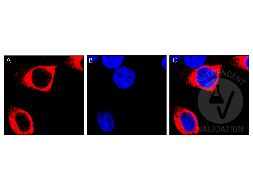
Staining with ABIN1537116 shows a perinuclear pattern, suggesting MR1 localizes in the endoplasmic reticulum. No signal was detected in sample negative control tissue and the secondary antibody only control.
Validation #101753 (Immunocytochemistry)
 Validation Images
![Human MR1-expressing HEK293 cells were stained with MR1 antibody ABIN1537116 and a Texas Red-conjugated secondary antibody (red, A). For nuclear staining, cells were stained with Hoechst (blue, B). C shows both channels merged.]() Human MR1-expressing HEK293 cells were stained with MR1 antibody ABIN1537116 and a Texas Red-conjugated secondary antibody (red, A). For nuclear staining, cells were stained with Hoechst (blue, B). C shows both channels merged.
Full Methods
Human MR1-expressing HEK293 cells were stained with MR1 antibody ABIN1537116 and a Texas Red-conjugated secondary antibody (red, A). For nuclear staining, cells were stained with Hoechst (blue, B). C shows both channels merged.
Full Methods -
- by
- Dr. Randy Brutkiewicz Laboratory, Department of Microbiology and Immunology, Indiana University School of Medicine
- No.
- #102828
- Date
- 02/20/2018
- Antigen
- MR1
- Lot Number
- SA111213CH
- Method validated
- Immunoprecipitation
- Positive Control
- HEK293 cells transfected with human MR1 cDNA
- Negative Control
- HEK293 cells transfected with plasmid vector only
- Notes
Passed. ABIN1537116 immunoprecipitates human MR1 overexpressed by HEK293 cells.
- Primary Antibody
- ABIN1537116
- Secondary Antibody
- goat anti-rabbit Dye-IR800 conjugated antibody (Advansta, R-05060-250, lot 17083179)
- Full Protocol
- Grow HEK293 cells in DMEM medium (Lonza, 12-614F, lot 0000618582) supplemented with serum (Hyclone, SH30071.03, lot AAG205460) and antibiotics (Hyclone, SV30010, lot J150013), at 37°C and 5% CO2 dish to 70-90% confluency.
- Transfect cells with pCDNA 3.1 neo (-) (Invitrogen) containing human MR1 cDNA (Genecopoeia) using Polyethylenimine (Polysciences, 23966) following the manufacturer´s instructions.
- Lyse cells in cold lysis buffer (10mM Tris pH7.4, 150mM NaCl, 0.5mM EDTA, 2% CHAPS).
- Determine total protein content of the lysates using Commassie Protein Assay Reagent (Thermo Scientific, 1856209, lot NL179252).
- Immobilize 100µl of protein G-conjugated Sepharose beads (Pierce, product 20399, lot RI239318) ON at 4°C with
- 2.5µg mouse anti-MR1 antibody (antibodies-online, ABIN2665876, lot B177559),
- 2.5µg mouse anti-MR1 antibody (antibodies-online, ABIN516526, lot12045-5B5),
- 2.5µg rabbit anti-MR1 antibody (antibodies-online, ABIN1537116, lot SA111213CH),
- 2.5µg mouse IgG2a antibody (Biolegend, 400202, lot B153642),
- 2.5µg mouse IgG1 antibody (BD, 555746, lot 3221830), or
- 2.5µg rabbit IgG antibody (Santa Cruz Biotechnology, SC-5560, lot E0609).
- Incubate 500µg of the cell lysates with 2.5µg of antibody-bead conjugate ON at 4°C.
- Wash lysates 4x with PBS.
- Denature beads for 5min at 95°C in 60µl Laemmli SDS sample buffer and subsequently separate them on a SDS-PAGE gel using Acrylamide/Bis Premixed (Bio-Rad, 61-0125, lot 260000477) for 2-3h at 100V.
- Transfer proteins onto PVDF membrane (Millipore, IPVH00010, lot K5AA6843U) with a Western blotting system for ON at 4°C at 150mA.
- Block the membrane with blocking buffer (2% BSA/PBS/0.05%Tween-20) for 1h at RT.
- Incubate membrane with primary rabbit anti-MR1 antibody (antibodies-online ABIN1537116, lot SA111213CH) diluted 1:1000 in blocking buffer ON at 4°C.
- Wash membrane 3x for 10min with PBS/0.05%Tween-20.
- Incubate membrane with secondary goat anti-rabbit Dye-IR800 conjugated antibody (Advansta, R-05060-250, lot 17083179) diluted 1:10000 in PBS/0.05% Tween-20 for 1h at RT.
- Wash membrane 3x for 10 min with PBS/0.05% Tween-20.
- Reveal protein bands using an Odyssey imaging system (LI-COR Biosciences).
- Experimental Notes
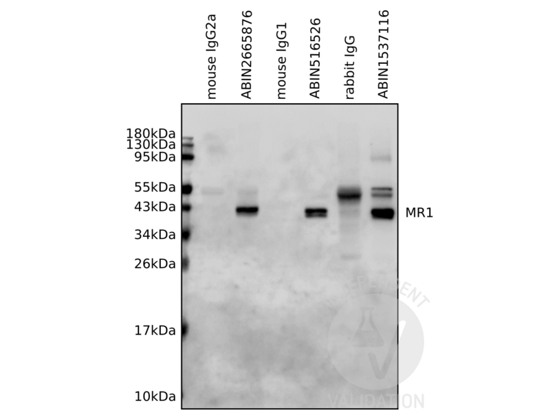
The human MR1 antibody ABIN1537116, but not the isotype control, immunoprecipitates with human MR1 overexpressed by HEK293 cells.
Validation #102828 (Immunoprecipitation)
 Validation Images
![Lysates from human MR1-expressing HEK293 cells were immunoprecipitated by antibodies specific for MR1 (ABIN2665876, ABIN516526, ABIN1537116) or the respective isotype controls (mouse IgG2a, mouse, IgG1, rabbit IgG). Immunoprecipitants were resolved by SDS-PAGE gel followed by Western blotting analysis using MR1 antibody ABIN1537116.]() Lysates from human MR1-expressing HEK293 cells were immunoprecipitated by antibodies specific for MR1 (ABIN2665876, ABIN516526, ABIN1537116) or the respective isotype controls (mouse IgG2a, mouse, IgG1, rabbit IgG). Immunoprecipitants were resolved by SDS-PAGE gel followed by Western blotting analysis using MR1 antibody ABIN1537116.
Full Methods
Lysates from human MR1-expressing HEK293 cells were immunoprecipitated by antibodies specific for MR1 (ABIN2665876, ABIN516526, ABIN1537116) or the respective isotype controls (mouse IgG2a, mouse, IgG1, rabbit IgG). Immunoprecipitants were resolved by SDS-PAGE gel followed by Western blotting analysis using MR1 antibody ABIN1537116.
Full Methods -
- by
- Dr. Randy Brutkiewicz Laboratory, Department of Microbiology and Immunology, Indiana University School of Medicine
- No.
- #102829
- Date
- 02/20/2018
- Antigen
- MR1
- Lot Number
- SA111213CH
- Method validated
- Western Blotting
- Positive Control
- HEK293 cells transfected with human MR1 cDNA
- Negative Control
- HEK293 cells transfected with plasmid vector only
- Notes
Passed. ABIN1537116 recognizes human MR1 overexpressed by HEK293 cells in a western blot.
- Primary Antibody
- ABIN1537116
- Secondary Antibody
- goat anti-rabbit Dye-IR800 conjugated antibody (Advansta, R-05060-250, lot 17083179)
- Full Protocol
- Grow HEK293 cells in DMEM medium (Lonza, 12-614F, lot 0000618582) supplemented with serum (Hyclone, SH30071.03, lot AAG205460) and antibiotics (Hyclone, SV30010, lot J150013), at 37°C and 5% CO2 dish to 70-90% confluency.
- Transfect cells with pCDNA 3.1 neo (-) (Invitrogen) containing human MR1 cDNA (Genecopoeia) using Polyethylenimine (Polysciences, 23966) following the manufacturer´s instructions.
- Lyse cells in cold lysis buffer (10mM Tris pH7.4, 150mM NaCl, 0.5mM EDTA, 2% CHAPS).
- Determine total protein content of the lysates using Commassie Protein Assay Reagent (Thermo Scientific, 1856209, lot NL179252).
- Denature 200µg total protein for 5min at 95°C in 20µl Laemmli SDS sample buffer and subsequently separate them on a SDS-PAGE gel using Acrylamide/Bis Premixed (Bio-Rad, 61-0125, lot 260000477) for 2-3h at 100V.
- Transfer proteins onto PVDF membrane (Millipore, IPVH00010, lot K5AA6843U) with a Western blotting system for ON at 4°C at 150mA.
- Block the membrane with blocking buffer (2% BSA/PBS/0.05%Tween-20) for 1h at RT.
- Incubate membrane with:
- primary rabbit anti-MR1 antibody (antibodies-online, ABIN1537116, lot SA111213CH) diluted 1:1000 in blocking buffer ON at 4°C.
- loading control rabbit anti beta-actin (antibodies-online, ABIN962807) diluted 1:500 in blocking buffer ON at 4°C.
- Wash membrane 3x for 10min with PBS/0.05%Tween-20.
- Incubate membrane with secondary goat anti-rabbit Dye-IR800 conjugated antibody (Advansta, R-05060-250, lot 17083179) diluted 1:10000 in PBS/0.05% Tween-20 for 1h at RT.
- Wash membrane 3x for 10min with PBS/0.05% Tween-20.
- Reveal protein bands using an Odyssey imaging system (LI-COR Biosciences).
- Experimental Notes
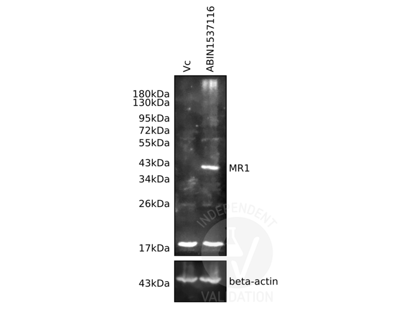
The human MR1 antibody ABIN1537116 reveals a protein of the expected molecular weight of MR1 in lysates of human MR1-expressing HEK293 cells. The protein bands is only visible in the positive but not the negative controls.
Validation #102829 (Western Blotting)
 Validation Images
![Lysates from human MR1-expressing HEK293 cells (MR1) and vector control cells (Vc) were resolved on a 10% SDS-PAGE gel for Western blotting analysis using antibodies specific for MR1 (ABIN1537116, upper panel) and beta-actin (ABIN962807, lower panel).]() Lysates from human MR1-expressing HEK293 cells (MR1) and vector control cells (Vc) were resolved on a 10% SDS-PAGE gel for Western blotting analysis using antibodies specific for MR1 (ABIN1537116, upper panel) and beta-actin (ABIN962807, lower panel).
Full Methods
Lysates from human MR1-expressing HEK293 cells (MR1) and vector control cells (Vc) were resolved on a 10% SDS-PAGE gel for Western blotting analysis using antibodies specific for MR1 (ABIN1537116, upper panel) and beta-actin (ABIN962807, lower panel).
Full Methods -
-
Format
- Liquid
-
Buffer
- Purified polyclonal antibody supplied in PBS with 0.09 % (W/V) sodium azide.
-
Preservative
- Sodium azide
-
Precaution of Use
- This product contains Sodium azide: a POISONOUS AND HAZARDOUS SUBSTANCE which should be handled by trained staff only.
-
Storage
- 4 °C,-20 °C
-
Storage Comment
- MR1 Antibody (C-term) can be refrigerated at 2-8 °C for up to 6 months. For long term storage, keep at -20 °C.
-
Expiry Date
- 6 months
-
-
- MR1 (Major Histocompatibility Complex, Class I-Related (MR1))
-
Alternative Name
- MR1
-
Background
- MR1 has antigen presentation function. Involved in the development and expansion of a small population of T cells expressing an invariant T cell receptor alpha chain called mucosal-associated invariant T cells (MAIT). MAIT cells are preferentially located in the gut lamina propria and therfore may be involed in monitoring commensal flora or serve as a distress signal. Expression and MAIT cell recognition seem to be ligand-dependent.
-
Molecular Weight
- 39366
-
Gene ID
- 3140
-
NCBI Accession
- NP_001181928, NP_001181929, NP_001181964, NP_001522
-
UniProt
- Q95460
-
Pathways
- Regulation of Leukocyte Mediated Immunity, Positive Regulation of Immune Effector Process, Production of Molecular Mediator of Immune Response, Cancer Immune Checkpoints
Target
-


