CALB1 antibody
 (2 validations)
Quick Overview for CALB1 antibody (ABIN1679483)
Target
See all CALB1 AntibodiesReactivity
Host
Clonality
Conjugate
Application
-
-
Cross-Reactivity
- Mouse, Rat
-
Characteristics
- Monoclonal Antibodies
-
Purification
- Affinity purification
-
Immunogen
- A synthesized peptide derived from human Calbindin
-
Isotype
- IgG
-
-
anti-Calbindin (CALB1) antibody
CALB1 Reactivity: Human IHC, WB, ELISA Host: Mouse Monoclonal 5A9 unconjugated
anti-Calbindin (CALB1) (AA 2-175) antibodyCALB1 Reactivity: Human, Rat, Mouse WB, IHC (p), FACS Host: Rabbit Polyclonal unconjugated
anti-Calbindin (CALB1) (Internal Region) antibodyVerified CALB1 Reactivity: Human, Rat IHC, WB, ELISA Host: Goat Polyclonal unconjugated
anti-Calbindin (CALB1) (AA 7-96) antibodyCALB1 Reactivity: Human IHC, ELISA, Coat, StM Host: Mouse Monoclonal CALB1-3333 unconjugated
anti-Calbindin (CALB1) (AA 2-175) antibodyCALB1 Reactivity: Human, Rat, Mouse IHC, WB, IP Host: Rabbit Polyclonal unconjugated
anti-Calbindin (CALB1) antibodyCALB1 Reactivity: Human, Rat, Mouse, Cow, Zebrafish (Danio rerio), Chicken, Grasshopper, Monkey, Non-Human Primate, Turtle IHC, WB, IP, ICC, IHC (p) Host: Rabbit Polyclonal unconjugated
anti-Calbindin (CALB1) (AA 7-96) antibodyCALB1 Reactivity: Human IHC Host: Mouse Monoclonal 471M unconjugated
anti-Calbindin (CALB1) (AA 1-261) antibodyCALB1 Reactivity: Human IHC, WB Host: Rabbit Polyclonal unconjugated
anti-Calbindin (CALB1) (C-Term) antibodyCALB1 Reactivity: Human, Rat, Mouse WB, ELISA, IHC (p) Host: Rabbit Polyclonal unconjugated
anti-Calbindin (CALB1) (AA 1-261) antibodyCALB1 Reactivity: Human WB, ELISA Host: Mouse Monoclonal 1F6 unconjugated
-
-
Application Notes
- WB,1:500 - 1:2000,IF,1:50 - 1:200
-
Restrictions
- For Research Use only
-
-
- by
- Prof. Merighi, Laboratory of Neurobiology, Department of Veterinary Sciences, University of Turin
- No.
- #104495
- Date
- 08/02/2023
- Antigen
- CALB1
- Lot Number
- 4000000953
- Method validated
- Immunohistochemistry
- Positive Control
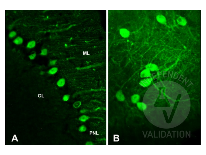
Adult (>2 months) CD1 mouse cerebellum (6 µm glass-mounted microtome sections)
Postnatal day 6-7 CD1 mouse cerebellum (cultured cerebellar slices)
- Negative Control
Control slices were processed for each experimental procedure, omitting the primary antibody; overnight incubation in diluent solution only.
- Notes
Passed. The CALB1 antibody ABIN3018777 works in IHC-P on cultured cerebellar slices at 1:50 dilution.
- Primary Antibody
- ABIN3018777
- Secondary Antibody
goat anti-mouse IgG (H+L) AF488-conjugated antibody (Thermo Fisher Scientific, A11034, lot 2380031)
- Full Protocol
- Indirect IMF on microtome sections
- Perfuse adult (>2 months) CD1 mice with paraformaldehyde 4% in 0.1 M phosphate buffer pH 7.4 and post-fix in the same fixative for an additional 2 h at RT.
- Wash, dehydrate, and embed samples in paraffin wax.
- Wash several times with 0.01 M PBS.
- Cut the cerebellum with a microtome into 6 µm sections and mount them on glass slides.
- After paraffin removal, incubate sections for 1 h at RT in PBS containing 1% albumin from chicken egg white (Sigma, A5378) and 0.3% Triton-X-100 (BioRad, 161-0407, lot 00583) to block non-specific binding sites.
- Incubate sections with primary mouse anti-CALB1 antibody (antibodies-online, ABIN3018777, lot 4000000953) diluted 1:50, 1:100, and 1:200 in 0.1 M PBS-BSA (Sigma, A7906)-PLL (Sigma, P1524) ON at RT.
- Wash 3x 5 min in 0.01 M PBS.
- Incubate sections with secondary goat anti-mouse IgG (H+L) AF488-conjugated antibody (Invitrogen by Thermo Fisher Scientific, A11034, lot 2380031) diluted 1:500 in 0.1 M PBS for 1 h at RT.
- Wash 3x 5 min in 0.01M PBS.
- Mount specimens in Fluoroshield (Sigma-Aldrich, F6182, lot MKCB0153V).
- Acquire Images with Leica DM 6000B fluorescence microscope equipped with a digital camera at 20-40x magnification.
- Indirect IMF on cultured cerebellar slices
- Euthanize CD1 mice at postnatal day 6-7 (P6-P7) with an overdose of 60 mg⁄100 g body weight sodium pentobarbital (Merck Life Science, Y0002194).
- Remove the brain removed from the skull while the head is kept submerged in ice-cooled Gey’s solution (Sigma-Aldrich) supplemented with glucose and antioxidants (for 500 mL: 4.8 mL 50% glucose, 0.05 g ascorbic acid, 0.1 g sodium pyruvate).
- Dissect the cerebellum from the brain.
- Cut 350 μm thick parasagittal slices of the cerebellum with a McIlwain tissue chopper (Brinkmann Instruments).
- Plate two to three slices onto a Millicell Cell Culture Insert (Merck Life Science, PICM0RG50).
- Place each insert inside a 35 mm Petri dish containing 1 mL of culture medium consisting of 50 % Eagle basal medium (BME, Sigma Chemicals), 25 % horse serum (Gibco by Thermo Fisher Scientific), 25 % Hanks balanced salt solution (Sigma-Aldrich), 0.5 % glucose, 0.5 % 200 mM L-glutamine, and 1ؘ % antibiotic/antimycotic solution.
- Incubate slices at 34 °C in 5 % CO2 for up to 20 d in vitro (DIV). Change the medium twice a week. Slices were allowed to equilibrate to the in vitro conditions for at least 4-6 DIV before IMF.
- Remove the culture medium from the dish and fix the slices in 1 mL of 4 % paraformaldehyde (Merck Life Science, P6148) in PBS for 1 h.
- Wash 3x 5 min in 0.01 M PBS.
- Incubate fixed cultures in PBS containing 1 % Triton X-100 (BioRad, 161-0407, lot 00583) for 10 min.
- Wash 3x 5 min in 0.01 M PBS.
- Incubate cultures ON at 4 °C under continuous stirring in PBS containing 1 % albumin from chicken egg white (Sigma, A5378) and 0.3 % Triton-X-100 (BioRad, 161-0407, lot 00583) to block non-specific binding sites.
- Incubate cultures with the primary mouse anti-CALB1 antibody (antibodies-online, ABIN3018777, lot 4000000953) diluted 1:50 in PBS-BSA (Sigma, A7906)-PLL (Sigma, P1524) ON at RT.
- Wash 5 x 5min in PBS.
- Incubate cultures with the secondary anti-rabbit antibody Alexa Fluor 488 diluted (Invitrogen by Thermo Fisher Scientific, A11034, lot 2380031) 1:500 in 0.1 M PBS for 1 h at RT.
- Wash 3x 5 min in 0.01 M PBS.
- Mount specimens in Fluoroshield (Sigma-Aldrich, F6182, lot MKCB0153V).
- Acquire Images with Leica DM 6000B fluorescence microscope equipped with a digital camera at 20-40x magnification.
- Experimental Notes
For indirect IMF on cerebellum paraffin sections, antigen retrieval treatment was also tested. In this case, sections were processed for microwave antigen retrieval for 10 min (95-100 °C) in 10 mM sodium citrate buffer (pH 6.0). After 20 min of spontaneous cooling, sections were washed twice for 5 min with distilled water and twice for 5 min with PBS.
Validation #104495 (Immunohistochemistry)
 Validation Images
![]() Full Methods
Full Methods -
- by
- Prof. Merighi, Laboratory of Neurobiology, Department of Veterinary Sciences, University of Turin
- No.
- #104531
- Date
- 08/02/2023
- Antigen
- CALB1
- Lot Number
- 4000000953
- Method validated
- Western Blotting
- Positive Control
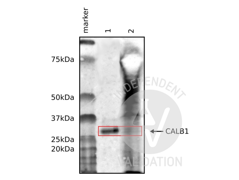
Adult mouse cerebellum and microdissected Purkinje neurons
- Negative Control
- Notes
Passed. The CALB1 antibody ABIN3018777 works in WB at a 1:1000 dilution with sensitive ECL substrate in adult mouse brain.
- Primary Antibody
- ABIN3018777
- Secondary Antibody
goat anti-rabbit IgG (H+L) HRP-conjugated (Thermo Fisher Scientific, G-21234)
- Full Protocol
- Homogenize tissues and neurons with cold lysis buffer containing 50 mM Tris HCl, 150 mM NaCl, 1% Triton X-100, 1 mM EDTA, and 1% protease inhibitor (Sigma P8340) using an ultrasonic homogenizer (MSE, SoniPrep 150) with 16 amplitude, 20 s on, 10 s off pulse for 60 s.
- Centrifuge tissue homogenates at 13,000 rpm for 20 min at 4 °C.
- Collect supernatants and determine total protein content using a Bradford assay.
- Denature 50 µg of total protein for 5 min at 90 °C and subsequently separate them on a denaturing 12% PAGE-SDS gel alongside a Precision Plus Protein Dual Color Standard (Bio-Rad, 160374).
- Electro-transfer proteins onto nitrocellulose membrane (Amersham Biosciences, RPN203D) ON in the cold room.
- Wash membrane 3x for 10 min with 0.01 M PBS containing 0.1% Tween-20 (PBST).
- Block membrane with PBST containing 2% bovine serum albumin for 1 h at RT.
- Incubate membrane with primary rabbit anti-CALB1 antibody (antibodies-online, ABIN3018777, lot 4000000953) diluted 1:1,000 in PBST ON at 4 °C.
- Wash membrane 3x 10 min with PBST.
- Incubate membrane with secondary HRP-conjugated goat anti-rabbit IgG (Thermo Fisher Scientific, G-21234) diluted 1:50,000 in PBST for 1 h at RT.
- Wash membrane 3x 10 min with PBST.
- Visualize proteins with SuperSignal West Atto Ultimate Sensitivity Substrate (Thermo Fisher Scientific, A38555) using a ChemiDoc Imaging System.
- Experimental Notes
Non-specific bands were also detected in adult mouse cerebellum tissue extracts, but not on laser-microdissected Purkinje neurons.
Validation #104531 (Western Blotting)
 Validation Images
![Western blot using ABIN3018777 to reveal CALB1 (28 kDa) in mouse laser-microdissected Purkinje neurons (1), and whole cerebellum (2).]() Western blot using ABIN3018777 to reveal CALB1 (28 kDa) in mouse laser-microdissected Purkinje neurons (1), and whole cerebellum (2).
Full Methods
Western blot using ABIN3018777 to reveal CALB1 (28 kDa) in mouse laser-microdissected Purkinje neurons (1), and whole cerebellum (2).
Full Methods -
-
Buffer
- PBS with 0.02 % sodium azide,0.05 % BSA,50 % glycerol, pH 7.3.
-
Preservative
- Sodium azide
-
Precaution of Use
- This product contains Sodium azide: a POISONOUS AND HAZARDOUS SUBSTANCE which should be handled by trained staff only.
-
Storage
- -20 °C
-
Storage Comment
- Store at -20°C. Avoid freeze / thaw cycles.
-
-
- CALB1 (Calbindin (CALB1))
-
Alternative Name
- CALB1
-
Background
- The protein encoded by this gene is a member of the calcium-binding protein superfamily that includes calmodulin and troponin C. Originally described as a 27 kDa protein, it is now known to be a 28 kDa protein. It contains four active calcium-binding domains, and has two modified domains that are thought to have lost their calcium binding capability. This protein is thought to buffer entry of calcium upon stimulation of glutamate receptors. Depletion of this protein was noted in patients with Huntington disease. [provided by RefSeq, Jan 2015],CALB, D-28K,Calcium Signaling,Cell Type Marker,Cell Type Marker_Neuron marker,Neuroscience,CALB1
-
Molecular Weight
- 30 kDa
-
Gene ID
- 793
-
UniProt
- P05937
Target
-


