IdU antibody
 (1 validation)
Quick Overview for IdU antibody (ABIN2669973)
Target
Reactivity
Host
Clonality
Conjugate
Application
Clone
-
-
Purification
- Purified from mouse ascites fluids by affinity chromatography
-
Immunogen
- Iododeoxyuridine coupled to keyhole limpet hemocyanin.
-
Isotype
- IgG2b
-
-
anti-Iododeoxyuridine (IdU) antibody
IdU Reactivity: Chemical IHC, IF Host: Mouse Monoclonal OTI7C9 unconjugated
anti-Iododeoxyuridine (IdU) antibodyIdU Reactivity: Chemical IHC, IF Host: Mouse Monoclonal OTI3F3 unconjugated
anti-Iododeoxyuridine (IdU) antibodyIdU Reactivity: Chemical IHC, WB, ELISA, FM, FACS Host: Mouse Monoclonal 32D8-D9 unconjugated
-
-
Application Notes
- IHC 1:150, IF 1:150,
-
Restrictions
- For Research Use only
-
-
- by
- Prof. Merighi, Laboratory of Neurobiology, Department of Veterinary Sciences, University of Turin
- No.
- #103750
- Date
- 04/19/2019
- Antigen
- IdU
- Lot Number
- W001
- Method validated
- Immunohistochemistry
- Positive Control
Adult (24 months) mouse brain and intestine (duodenum)
- Negative Control
We incubated slices overnight with the blocking solution only and then processed them with the secondary antibody.
- Notes
Passed. ABIN2669973 works in IHC-P with a microwave antigen retrieval pretreatment and concentration of 1:50.
- Primary Antibody
- ABIN2669973
- Secondary Antibody
- goat anti-mouse AF594-conjugated (Invitrogen by Thermo Fisher Scientific, A11032, lot 819561)
- Full Protocol
- Inject mouse intraperitoneally at the dose of 0.057mg/g body weight with IdU (Sigma, I7125) dissolved in sterile distilled water at final concentration of 100mg/ml, pH7.5.
- Perfuse mouse with 4% paraformaldehyde in 0.1M phosphate buffer (PB) pH7.4 and post-fix samples in the same fixative for an additional 2h at RT.
- Wash, dehydrate, and embed samples in paraffin wax.
- Following several washes in PBS, cut intestines and brain with a microtome (6µm-thick sections) and mount sections on glass slides.
- After paraffin removal, process sections by microwave antigen retrieval for 10min (95-100°C) in 10mM sodium citrate buffer pH6.0.
- Let sections cool for 20min.
- Wash sections 2x 5min with distilled water.
- Wash sections 5min with PBS.
- Incubate sections in PBS containing 1% albumin from chicken egg white (Sigma, A5378) and 0.3% Triton-X-100 (BioRad, 161-0407, lot 00583) for 1h at RT to block non-specific binding sites.
- Incubate sections with primary antibody mouse anti-IdU (antibodies-online, ABIN2669973, lot W001) diluted 1:50 in PBS-BSA-PLL ON at 4°C.
- Wash sections 3x 5min with 0.01M PBS.
- Incubate sections with secondary goat anti-mouse AF594-conjugated (Invitrogen by Thermo Fisher Scientific, A11032, lot 819561) diluted 1:500 in 0.1M PBS for 1h at RT.
- Wash sections 3x 5min with 0.01M PBS.
- Mount sections in Fluoroshield (Sigma, F6182, lot MKCB0153V).
- Acquire images with Leica DM 6000B fluorescence microscope equipped with the manufacturer’s FITC filter set and digital camera at magnifications of 20x and 40x. Parameters for image acquisition (Exposure 2.2s, Gain 2.3x, Saturation 1.5, Gamma 1.10) were maintained unchanged for all images.
- Experimental Notes
For the incubation with the primary antibody ABIN2669973 and 1:20, 1:70, and 1:100 dilutions were also tested with 0.01M PBS, 5% Normal Goat Serum (NGS Sigma, G9023, lot SLBV1396) and 0.1% Triton-X -100 as the blocking solution, but 1:50 and PBS containing 1% albumin from chicken egg white and 0.3% Triton-X gave the best results.
Validation #103750 (Immunohistochemistry)
 Validation Images
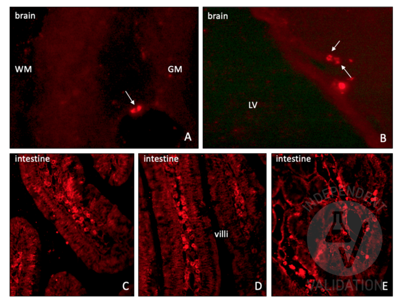
![Staining of mouse brain and intestine sections using ABIN2669973. A-B: A bright staining in the nuclei of individual cells scattered across the brain is present, especially in the sub-cortical areas at the border between the gray and white matters (A) and ventricular wall (B). Abbreviations: GM = gray matter; LV = lateral ventricle; WM = white matter. C-E: positive control staining in the intestine (duodenum). IdU+ proliferating cells are clearly detected in the germinal layer of the intestinal epithelium, at the basis of the duodenal villi.]() Staining of mouse brain and intestine sections using ABIN2669973. A-B: A bright staining in the nuclei of individual cells scattered across the brain is present, especially in the sub-cortical areas at the border between the gray and white matters (A) and ventricular wall (B). Abbreviations: GM = gray matter; LV = lateral ventricle; WM = white matter. C-E: positive control staining in the intestine (duodenum). IdU+ proliferating cells are clearly detected in the germinal layer of the intestinal epithelium, at the basis of the duodenal villi.
Full Methods
Staining of mouse brain and intestine sections using ABIN2669973. A-B: A bright staining in the nuclei of individual cells scattered across the brain is present, especially in the sub-cortical areas at the border between the gray and white matters (A) and ventricular wall (B). Abbreviations: GM = gray matter; LV = lateral ventricle; WM = white matter. C-E: positive control staining in the intestine (duodenum). IdU+ proliferating cells are clearly detected in the germinal layer of the intestinal epithelium, at the basis of the duodenal villi.
Full Methods -
-
Format
- Liquid
-
Concentration
- 4.54 mg/mL
-
-
- IdU (Iododeoxyuridine (IdU))
-
Alternative Name
- IdU
Target
-


