Recombinant RAD21 antibody (N-Term)
 (1 reference)
 (1 validation)
Quick Overview for Recombinant RAD21 antibody (N-Term) (ABIN6731038)
Target
See all RAD21 AntibodiesAntibody Type
Reactivity
Host
Clonality
Conjugate
Application
-
-
Binding Specificity
- N-Term
-
Brand
- AbFlex®
-
Purification
- Protein A Chromatography
-
Immunogen
- This antibody was raised against recombinant protein corresponding to the N-terminal half of mouse Rad21.
-
Isotype
- IgG2a
-
-
anti-RAD21 Homolog (RAD21) (Center) antibody
RAD21 Reactivity: Human, Mouse WB, IF, IP, IHC (p), ICC Host: Rabbit Polyclonal unconjugated
anti-RAD21 Homolog (RAD21) antibodyRAD21 Reactivity: Human WB, IHC, IF, IP, ChIP Host: Rabbit Polyclonal unconjugated
anti-RAD21 Homolog (RAD21) (AA 287-403) antibodyRAD21 Reactivity: Human, Rat, Monkey WB, ELISA, IHC, FACS, ICC Host: Mouse Monoclonal 1B6D1 unconjugated
anti-RAD21 Homolog (RAD21) antibodyKD Validated RAD21 Reactivity: Human WB, FACS, ICC Host: Rabbit Monoclonal 24GB1390 unconjugated Recombinant Antibody
anti-RAD21 Homolog (RAD21) (C-Term) antibodyRAD21 Reactivity: Human, Mouse WB, ELISA, IHC, IF, ICC Host: Rabbit Polyclonal unconjugated
anti-RAD21 Homolog (RAD21) antibodyRAD21 Reactivity: Human WB, ChIP Host: Rabbit Monoclonal unconjugated
anti-RAD21 Homolog (RAD21) (AA 373-422) antibodyRAD21 Reactivity: Human, Mouse, Rat, Cow, Rabbit, Dog, Horse, Monkey, Guinea Pig, Chicken, Bat, Pig, Xenopus laevis WB Host: Rabbit Polyclonal unconjugated
anti-RAD21 Homolog (RAD21) (C-Term) antibodyRAD21 Reactivity: Human, Mouse, Rat, Cow, Zebrafish (Danio rerio) WB, IHC, IF, IC Host: Rabbit Polyclonal unconjugated
anti-RAD21 Homolog (RAD21) (C-Term) antibodyRAD21 Reactivity: Human, Mouse, Hamster WB, IF Host: Rabbit Polyclonal unconjugated
anti-RAD21 Homolog (RAD21) (AA 531-631) antibodyRAD21 Reactivity: Human, Rat WB, ELISA, FACS, IF (cc), IF (p), IHC (p), IHC (fro) Host: Rabbit Polyclonal unconjugated
-
-
Application Notes
-
Validated Applications:
IF: 0.25 - 1 µg/ml
WB: 0.5- 2 μg/mL
-
Comment
-
AbFlex® antibodies are recombinant antibodies (rAbs) that have been generated using defined DNA sequences to produce highly specific, reproducible antibodies. Each AbFlex antibody contains a 6XHis tag, an avidin tag sequence for enzymatic biotin conjugation using the biotin ligase, BirA, and a sortase recognition motif (LPXTG) to attach a variety of labels directly to the antibody including fluorophores, enzymatic substrates (HRP, AP), peptides, drugs as well as solid supports. AbFlex Rad21 antibody was expressed as full-length IgG with mouse immunoglobulin heavy and light chains (IgG2a isotype) in mammalian 293 cells
-
Restrictions
- For Research Use only
-
-
- by
- Cantù Lab, Gene Regulation during Development and Disease, Linköping University
- No.
- #104617
- Date
- 12/06/2024
- Antigen
- RAD21
- Lot Number
- Method validated
- Cleavage Under Targets and Release Using Nuclease
- Positive Control
- Anti H3K4me (antibodies-online, ABIN3023251)
- Negative Control
- Guinea Pig anti-rabbit IgG (antibodies-online, ABIN101961)
- Notes
Passed.
- Primary Antibody
- ABIN6731038
- Secondary Antibody
- Full Protocol
- Cell harvest and nuclear extraction
- Harvest 250,000 HEK293T cells per antibody
- Centrifuge cell solution 5 min at 600 x g at RT.
- Remove the liquid carefully.
- Gently resuspend cells in 2 mL of Nuclear Extraction Buffer (20 mM HEPES-KOH pH 8.2, 20% Glycerol, 0,05% IGEPAL, 0.5 mM Spermidine, 10 mM KCl, Roche Complete Protease Inhibitor EDTA-free).
- Move the solution to a 2 mL centrifuge tube.
- Pellet the nuclei 800 x g for 5 min.
- Repeat the NE wash twice for a total of three washes.
- Resuspend the nuclei in 40 µL NE Buffer per sample.
- Concanavalin A beads preparation
- Prepare one 2 mL microcentrifuge tube.
- Gently resuspend the magnetic Concanavalin A Beads (antibodies-online, ABIN6923139).
- Pipette 10 µL Con A Beads slurry for each sample into the 2 mL microcentrifuge tube.
- Place the tube on a magnet stand until the fluid is clear. Remove the liquid carefully.
- Remove the microcentrifuge tube from the magnetic stand.
- Pipette 1 mL Binding Buffer (20 mM HEPES pH 7.5, 10 mM KCl, 1 mM CaCl2, 1 mM MnCl2) into the tube and resuspend ConA beads by gentle pipetting.
- Spin down the liquid from the lid with a quick pulse in a table-top centrifuge.
- Place the tubes on a magnet stand until the fluid is clear. Remove the liquid carefully.
- Remove the microcentrifuge tube from the magnetic stand.
- Repeat the wash twice for a total of three washes.
- Gently resuspend the ConA Beads in 44ul binding buffer
- Nuclei immobilization – binding to Concanavalin A beads
- Carefully vortex the nuclei suspension and add 44 µL of the Con A beads in Binding Buffer to the cell suspension for each sample.
- Close tube tightly incubates 10 min at 4 °C.
- Put the 2 mL tube on the magnet stand and when the liquid is clear remove the supernatant.
- Resuspend the beads in 1 mL of EDTA wash buffer (20 mM HEPES pH 7.5, 150 mM NaCl, 0.5 mM Spermidine, Roche Complete Protease Inhibitor EDTA-free, 2mM EDTA)
- Incubate 5 min at RT.
- Place the tube on the magnet stand and when the liquid is clear remove the supernatant.
- Resuspend the beads in 200µl of wash buffer (20 mM HEPES pH 7.5, 150 mM NaCl, 0.5 mM Spermidine, Roche Complete Protease Inhibitor EDTA-free) x each sample.
- Primary antibody binding
- Divide nuclei suspension into separate 200 µL PCR tubes, one for each antibody.
- Add 2 µL antibody (RAD21 ABIN6731038, anti-H3K4me positive control ABIN3023251, and guinea pig anti-rabbit IgG negative control antibody ABIN101961) to the respective tube, corresponding to a 1:100 dilution.
- Incubate at 4 °C ON.
- Place the tubes on a magnet stand until the fluid is clear. Remove the liquid carefully.
- Remove the microcentrifuge tubes from the magnetic stand.
- Wash with 200 µL of Wash Buffer using a multichannel pipette to accelerate the process.
- Repeat the wash five times for a total of six washes.
- pAG-MNase Binding
- Prepare a 1.5 mL microcentrifuge tube containing 200 µL of pAG mix per sample (200 µL of wash buffer + 120 ng pAG-MNase per sample)
- Place the PCR tubes with the sample on a magnet stand until the fluid is clear. Remove the liquid carefully.
- Remove tubes from the magnetic stand.
- Resuspend the beads in 200 µL of pAG-MNase premix.
- Incubate 30 min at 4 °C
- Place the tubes on a magnet stand until the fluid is clear. Remove the liquid carefully.
- Remove the microcentrifuge tubes from the magnetic stand.
- Wash with 200 µL of Wash Buffer using a multichannel pipette to accelerate the process.
- Repeat the wash five times for a total of six washes.
- Resuspend in 100 µL of Wash Buffer.
- MNase digestion and release of pAG-MNase-antibody-chromatin complexes
- Place PCR tubes on ice and allow to chill.
- Prepare a 1.5 mL microcentrifuge tube with 102 µl of 2 mM CaCl2 mix per sample (100 µl Wash Buffer + 2 µL 100 mM CaCl2) and let it chill on ice.
- Always in ice, place the samples on the magnetic rack and when the liquid is clear remove the supernatant.
- Resuspend the samples in 100 µl of the 2 mM CaCl2 mix and incubate in ice for exactly 30 min.
- Place the sample on the magnet stand and when the liquid is clear remove the supernatant.
- Resuspend the sample in 50 µl of 1x Urea STOP Buffer (8.5 M Urea, 100 mM NaCl, 2 mM EGTA, 2 mM EDTA, 0,5% IGEPAL).
- Incubate the samples 1h at 4°C.
- Transfer the supernatant containing the pAG-MNase-bound digested chromatin fragments to fresh 200 µl PCR tubes.
- DNA Clean up (Mag-Bind® TotalPure NGS - M1378-01)
- Take the Mag-Bind® TotalPure NGS beads (Omega Bio-Tek, M1378-01) from the storage and wait until they are RT.
- Add 2x volume of beads to each sample (e.g. 100 µL of beads for 50 µL of sample).
- Incubate the beads and the sample for 15 min at RT.
- During incubation prepare fresh EtOH 80%.
- Place the PCR tubes on a magnet stand and when the liquid is clear remove the supernatant.
- Add 200 µl of fresh 80% EtOH to the sample without disturbing the beads (Important!!! Do NOT resuspend the beads or remove the tubes from the magnet stand or the sample will be lost).
- Incubate 30 sec at RT.
- Remove the EtOH from the sample.
- Repeat the wash with 80% EtOH.
- Resuspend the beads in 25 µL of 10 mM Tris.
- Incubate the sample for 2 min at RT.
- Repeat the 2x beads clean up as described before (this time with 50 µlL of beads for each sample).
- Resuspend the beads + DNA in 20 µL of 10 mM Tris.
- Library preparation and sequencing
- Prepare Libraries using KAPA HyperPrep Kit using KAPA Dual-Indexed adapters according to protocol.
- Sequence samples on an Illumina NextSeq 500 sequencer, using a NextSeq 500/550 High Output Kit v2.5 (75 Cycles), 36bp PE.
- Peak calling
- Trim reads using using bbTools bbduk (BBMap - Bushnell B. - sourceforge.net/projects/bbmap/) to remove adapters, artifacts and repeat sequences.
- Aligned reads were mapped to the hg38 human genome using bowtie with options -m 1 -v 0 -I 0 -X 500.
- Use SAMtools to convert SAM files to BAM files and remove duplicates.
- Use BEDtools genomecov to produce Bedgraph files.
- Call peaks using SEACR with a 0.001 threshold and the option norm stringent.
- Cell harvest and nuclear extraction
- Experimental Notes
Validation #104617 (Cleavage Under Targets and Release Using Nuclease)
 Validation Images
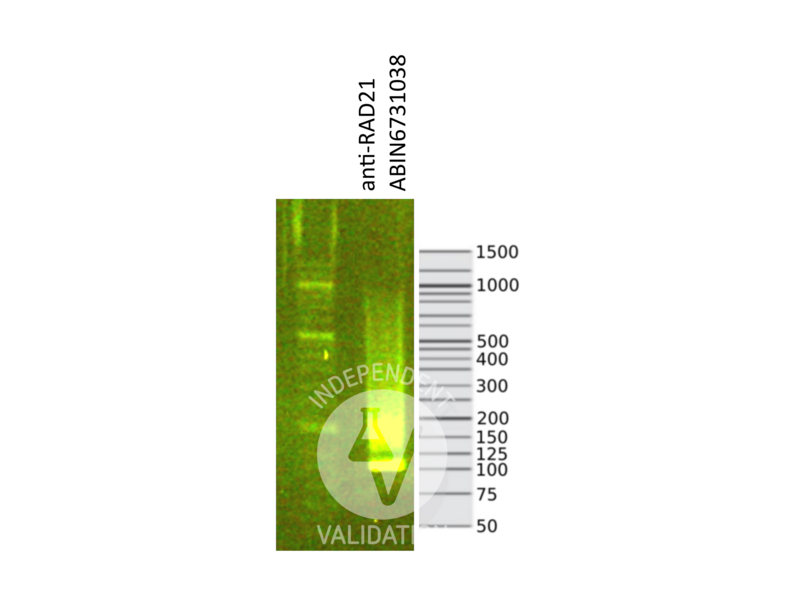
![Library profiles comparing fragment size distributions on an E-Gel EX 2% agarose gel (Thermo Fisher). Fragments obtained from CUT&RUN using RAD21 ABIN6731038 (right) after library preparation, compared to the E-Gel Sizing DNA Ladder (Thermo Fisher) (left).]() Library profiles comparing fragment size distributions on an E-Gel EX 2% agarose gel (Thermo Fisher). Fragments obtained from CUT&RUN using RAD21 ABIN6731038 (right) after library preparation, compared to the E-Gel Sizing DNA Ladder (Thermo Fisher) (left).
Library profiles comparing fragment size distributions on an E-Gel EX 2% agarose gel (Thermo Fisher). Fragments obtained from CUT&RUN using RAD21 ABIN6731038 (right) after library preparation, compared to the E-Gel Sizing DNA Ladder (Thermo Fisher) (left).
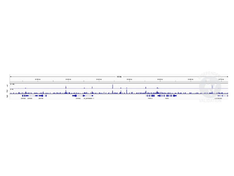
![1. Alignment tracks from CTCF CUT&RUN in HEK293T cells. 2. Alignment tracks from CUT&RUN targeting RAD21 in HEK293T cells using ABIN6731038 antibody showing the FBRS locus. 3. RefSeq Genes.]() 1. Alignment tracks from CTCF CUT&RUN in HEK293T cells. 2. Alignment tracks from CUT&RUN targeting RAD21 in HEK293T cells using ABIN6731038 antibody showing the FBRS locus. 3. RefSeq Genes.
Full Methods
1. Alignment tracks from CTCF CUT&RUN in HEK293T cells. 2. Alignment tracks from CUT&RUN targeting RAD21 in HEK293T cells using ABIN6731038 antibody showing the FBRS locus. 3. RefSeq Genes.
Full Methods -
-
Format
- Liquid
-
Buffer
- Purified IgG in 140 mM Hepes, pH 7.5, 70 mM NaCl, 32 mM NaOAc, 0.035 % sodium azide, 30 % glycerol.
-
Preservative
- Sodium azide
-
Precaution of Use
- This product contains Sodium azide: a POISONOUS AND HAZARDOUS SUBSTANCE which should be handled by trained staff only.
-
Storage
- -20 °C
-
-
-
: "Wnt signaling activation induces CTCF binding and loop formation at cis-regulatory elements of target genes." in: Genome research, Vol. 35, Issue 8, pp. 1701-1716, (2025) (PubMed).
-
: "Wnt signaling activation induces CTCF binding and loop formation at cis-regulatory elements of target genes." in: Genome research, Vol. 35, Issue 8, pp. 1701-1716, (2025) (PubMed).
-
- RAD21 (RAD21 Homolog (RAD21))
-
Alternative Name
- Rad21
-
Molecular Weight
- 100 kDa
-
Pathways
- Positive Regulation of Endopeptidase Activity
Target
-


