MYO1F antibody (AA 491-767)
 (2 validations)
Quick Overview for MYO1F antibody (AA 491-767) (ABIN7429520)
Target
See all MYO1F AntibodiesReactivity
Host
Clonality
Conjugate
Application
-
-
Binding Specificity
- AA 491-767
-
Purpose
- Polyclonal Antibody to Myosin IF (MYO1F)
-
Specificity
- The antibody is a rabbit polyclonal antibody raised against MYO1F. It has been selected for its ability to recognize MYO1F in immunohistochemical staining and western blotting.
-
Cross-Reactivity
- Mouse, Rat
-
Purification
- Antigen-specific affinity chromatography followed by Protein A affinity chromatography
-
Immunogen
- Recombinant Myosin IF (MYO1F) corresdonding to Glu491~Ser767 (Accession # O00160)
-
Isotype
- IgG
-
-
anti-Myosin IF (MYO1F) (AA 655-922) antibody
MYO1F Reactivity: Rat WB, IHC, IP, ICC Host: Rabbit Polyclonal unconjugated
anti-Myosin IF (MYO1F) (Internal Region) antibodyMYO1F Reactivity: Human, Mouse, Rat WB, ELISA Host: Rabbit Polyclonal unconjugated
-
-
Application Notes
- Western blotting: 0.01-2 μg/mL,Immunohistochemistry: 5-20 μg/mL,Immunocytochemistry: 5-20 μg/mL,Optimal working dilutions must be determined by end user.
-
Comment
-
The thermal stability is described by the loss rate. The loss rate was determined by accelerated thermal degradation test, that is, incubate the protein at 37°C for 48h, and no obvious degradation and precipitation were observed. The loss rate is less than 5% within the expiration date under appropriate storage condition.
-
Restrictions
- For Research Use only
-
-
- by
Zoe Linke, Lena Cook, Jörg W. Bartsch
Department of Neurosurgery, Philipps-University Marburg
- No.
- #104488
- Date
- 09/06/2023
- Antigen
- MYO1F
- Lot Number
- A20230217043
- Method validated
- Immunohistochemistry
- Positive Control
Pathologically altered human brain tissue
- Negative Control
Tissue samples processed without primary antibody
- Notes
Passed. ABIN6997802 specifically stains sub-membranous regions in brain tissue metastatic cells. in lysates of bone marrow derived mouse macrophages, indicating a distinct localization of MYO1F.
- Primary Antibody
- ABIN6997802
- Secondary Antibody
- VECTASTAIN® Elite® ABC-HRP Kit, Anti-Rabbit biotinylated antibody (Vector Laboratories, PK-6101, lot ZF0103)
- Full Protocol
- Incubate paraffin-embedded human brain metastases tissue sections on glass slides for 45 min at 60 °C.
- Deparaffinize and rehydrate sections in a descending xylene and graded alcohol series:
- xylene 2x for 10 min.
- 100 % (v/v) ethanol 2x for 5 min.
- 96 % (v/v) ethanol 2x for 5 min.
- 70 % (v/v) ethanol 2x for 5 min.
- Wash slides 1x 5 min with dH2O.
- Wash slides 2x 5 min with 1x PBS.
- Cook slides for 20 min at 120 °C in 600 mL citrate buffer pH 6 in a large glass cuvette.
- Cool down slides for 30 min at RT.
- Incubate slides in 1 % (v/v) H2O2 for 30 min to quench endogenous peroxidase.
- Wash slides 2x 5 min in 1x PBS.
- Block slides in blocking serum (1.5 % (v/v), 1X PBS; VECTASTAIN® Elite® ABC-HRP Kit, Peroxidase (Rabbit IgG), Vector Laboratories, PK-6101, lot ZF0103) for 30 min at RT.
- Incubate slides with primary rabbit anti-MYO1F antibody (antibodies-online, ABIN6997802, lot A20230217043) diluted 1:1,000 in blocking serum ON at 4 °C. Incubate the negative control with blocking serum only.
- Prepare ABC reagent from 2 % (v/v) solution A and 20 % (v/v) solution B and filled up with 1x PBS according to the manual. Incubate solution for 30 min at RT.
- Wash slides 2x 5 min in 1x PBS.
- Cover slides with 200 µL secondary antibody (VECTASTAIN® Elite® ABC-HRP Kit, Anti-Rabbit biotinylated antibody, Vector Laboratories, PK-6101, lot ZF0103) diluted 1:200 in 1.5 % (v/v) goat serum in 1x PBS and incubate for 30 min at RT.
- Wash slides 2x 5 min in 1x PBS.
- Cover slides with 200 µL ABC reagent and incubate for 30 min at RT.
- Wash slides 2x 5 min in 1X PBS.
- Prepare staining solution by diluting 3 % ImmPACT DAB Chromogen concentrate in ImmPACT DAB diluent (ImmPACT® DAB Substrate Kit, Peroxidase, Vector Laboratories, SK-4105, lot ZH0708) according to the manual.
- Apply 200 μL of the staining solution to the slides for 2.5 min.
- Immerse slides 2x for 5 min in 1X PBS to stop the staining reaction.
- Counterstain slides with hematoxylin (Roth, T865.2, lot 420302035) for 5 min.
- Wash slides for 15 min under running tap water.
- Dehydrate sections in an ascending alcohol-xylene series:
- 70% ethanol 2x for 5 min.
- 96% ethanol 2x for 5 min.
- 100% ethanol 2x for 5 min.
- Xylene 2x for 10 min.
- Mount sections with EUKITT® Quick-hardening mounting medium (Sigma-Aldrich, 0389-100ML, lot BCCC8832) according to the manual and cover with glass coverslips.
- Dry sections for 24 h.
- Image acquisition on Aperio® AT2 (Leica). Magnification 10x and 40x.
- Experimental Notes
Validation #104488 (Immunohistochemistry)
 Validation Images
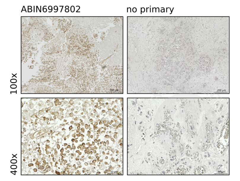
![IHC staining of human brain tissue for tumor cells with anti-MYO1F antibody ABIN6997802 (1:1,000) and counterstain with hematoxylin (left panels), highlighting the sub-membranous regions in the cells. Panels on the right show images obtained without primary antibody. Magnification 100x (upper panels) and 400x (lower panels).]() IHC staining of human brain tissue for tumor cells with anti-MYO1F antibody ABIN6997802 (1:1,000) and counterstain with hematoxylin (left panels), highlighting the sub-membranous regions in the cells. Panels on the right show images obtained without primary antibody. Magnification 100x (upper panels) and 400x (lower panels).
Full Methods
IHC staining of human brain tissue for tumor cells with anti-MYO1F antibody ABIN6997802 (1:1,000) and counterstain with hematoxylin (left panels), highlighting the sub-membranous regions in the cells. Panels on the right show images obtained without primary antibody. Magnification 100x (upper panels) and 400x (lower panels).
Full Methods -
- by
Zoe Linke, Lena Cook, Jörg W. Bartsch
Department of Neurosurgery, Philipps-University Marburg
- No.
- #104540
- Date
- 09/06/2023
- Antigen
- MYO1F
- Lot Number
- A20230217043
- Method validated
- Immunofluorescence
- Positive Control
Cultivated human breast cancer cells
- Negative Control
No primary antibody control
- Notes
Passed. ABIN6997802 specifically stains MYO1F in cultivated human breast cancer cells.
- Primary Antibody
- ABIN6997802
- Secondary Antibody
- donkey anti-rabbit IgG H&L (Alexa Fluor® 488) secondary antibody (Abcam, ab96919, lot GR43917-2)
- Full Protocol
- Incubate glass coverslips in a 24-well plate with 300 µL collagen I dilutes 1:20 with 10 mM HCl for each well for 1 h at 37 °C.
- Wash coverslips in well 2x with 500 µL 1X PBS.
- Seed 100,000 cells in 500 µL per well onto coverslips for 24 h at 37 °C and 5% CO2.
- Wash cells on coverslips 3x with 500 µL 1X PBS.
- Fix cells with 4 % (w/v) PFA in 1X PBS for 15 min.
- Permeabilize cells with 0.3 % (v/v) Triton X-100 in PBS for 15 min.
- Wash cells on coverslips 3x with 500 µL 1X PBS.
- Block cells with blocking solution (5 % (w/v) BSA in 1X PBS) for 1 h.
- Remove blocking solution and add primary rabbit anti-MYO1F antibody (antibodies-online, ABIN6997802, lot A20230217043) diluted 1:100 in blocking solution. Omit primary antibody for negative control.
- Incubate plate ON at 4 °C.
- Wash cells on coverslips 3x with 500 µL 1X PBS.
- Add donkey anti-rabbit IgG H&L (Alexa Fluor® 488) secondary antibody (Abcam, ab96919, lot GR43917-2) diluted 1:1000 in 5 % (w/v) BSA in 1X PBS.
- Incubate plate for 1 h at RT in the dark.
- Wash cells on coverslips 3x with 500 µL 1x PBS.
- Incubate cells on coverslips with Hoechst 33258 (Sigma-Aldrich, 861405)) diluted 1:10,000 in 1X PBS for 20 min at RT to stain nuclei.
- Wash cells on coverslips 3x with 500 µL 1X PBS.
- Mount coverslips with fluorescence mounting medium (Agilent, S302380-2, lot 11407565) and store at 4 °C in the dark.
- Acquire images with a confocal laser scanning microscope (Leica SP8i) with 63x magnification and process the images with ImageJ Fiji.
- Experimental Notes
Validation #104540 (Immunofluorescence)
 Validation Images
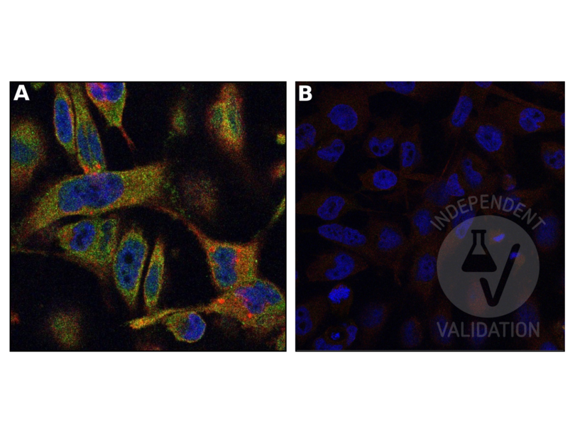
![A. IF staining in human cultivated breast cancer cells with anti-MYO1F antibody ABIN6997802 (1:100) and secondary antibody (1:1,000) labeled by green fluorescence and anti-ADAM8 antibody (1:100, product #PA5-47047, Invitrogen, lot #WL3451215) and Donkey anti-Goat IgG (H+L) Secondary Antibody, Texas Red (1:1,000, product #PA1-28662, Invitrogen, lot# VI3078250F) Nuclei stained with Hoechst 33258 (blue). MYO1F staining is observed in in vesicle like structures localized perinuclear and on the inner side of the cell membrane. Magnification 63x. B. Negative control without primary antibody.]() A. IF staining in human cultivated breast cancer cells with anti-MYO1F antibody ABIN6997802 (1:100) and secondary antibody (1:1,000) labeled by green fluorescence and anti-ADAM8 antibody (1:100, product #PA5-47047, Invitrogen, lot #WL3451215) and Donkey anti-Goat IgG (H+L) Secondary Antibody, Texas Red (1:1,000, product #PA1-28662, Invitrogen, lot# VI3078250F) Nuclei stained with Hoechst 33258 (blue). MYO1F staining is observed in in vesicle like structures localized perinuclear and on the inner side of the cell membrane. Magnification 63x. B. Negative control without primary antibody.
Full Methods
A. IF staining in human cultivated breast cancer cells with anti-MYO1F antibody ABIN6997802 (1:100) and secondary antibody (1:1,000) labeled by green fluorescence and anti-ADAM8 antibody (1:100, product #PA5-47047, Invitrogen, lot #WL3451215) and Donkey anti-Goat IgG (H+L) Secondary Antibody, Texas Red (1:1,000, product #PA1-28662, Invitrogen, lot# VI3078250F) Nuclei stained with Hoechst 33258 (blue). MYO1F staining is observed in in vesicle like structures localized perinuclear and on the inner side of the cell membrane. Magnification 63x. B. Negative control without primary antibody.
Full Methods -
-
Format
- Liquid
-
Concentration
- 0.5 mg/mL
-
Buffer
- PBS, pH 7.4, containing 0.02 % Sodium azide, 50 % glycerol.
-
Preservative
- Sodium azide
-
Precaution of Use
- This product contains Sodium azide: a POISONOUS AND HAZARDOUS SUBSTANCE which should be handled by trained staff only.
-
Storage
- 4 °C,-20 °C
-
Storage Comment
- Store at 4°C for frequent use. Stored at -20°C in a manual defrost freezer for two year without detectable loss of activity. Avoid repeated freeze-thaw cycles.
-
Expiry Date
- 24 months
-
-
- MYO1F (Myosin IF (MYO1F))
-
Alternative Name
- MYO1F
-
Background
- Myosin-Ie
-
UniProt
- O00160
Target
-


