Tissue factor antibody (AA 32-100)
 (7 references)
 (1 validation)
Quick Overview for Tissue factor antibody (AA 32-100) (ABIN708086)
Target
See all Tissue factor (F3) AntibodiesReactivity
Host
Clonality
Conjugate
Application
-
-
Binding Specificity
- AA 32-100
-
Cross-Reactivity
- Human, Mouse, Pig, Rat
-
Predicted Reactivity
- Dog,Cow,Horse,Rabbit,Guinea Pig
-
Purification
- Purified by Protein A.
-
Immunogen
- KLH conjugated synthetic peptide derived from human CD142
-
Isotype
- IgG
-
-
anti-Coagulation Factor III (thromboplastin, Tissue Factor) (F3) (AA 45-154) antibody
F3 Reactivity: Human WB, ELISA, IP, PLA Host: Mouse Monoclonal 4G4 unconjugated
anti-Coagulation Factor III (thromboplastin, Tissue Factor) (F3) antibodyF3 Reactivity: Human WB, ICC, FACS, IHC (p) Host: Mouse Monoclonal HTF-1 unconjugated
anti-Coagulation Factor III (thromboplastin, Tissue Factor) (F3) (AA 36-292) antibodyF3 Reactivity: Cow WB, IHC, ICC, IP Host: Rabbit Polyclonal unconjugated
anti-Coagulation Factor III (thromboplastin, Tissue Factor) (F3) (AA 45-154) antibodyF3 Reactivity: Human WB, ELISA Host: Mouse Monoclonal 4G4 unconjugated
anti-Coagulation Factor III (thromboplastin, Tissue Factor) (F3) (AA 1-295) antibodyF3 Reactivity: Human WB, FACS Host: Mouse Polyclonal unconjugated
anti-Coagulation Factor III (thromboplastin, Tissue Factor) (F3) (AA 1-295) antibodyF3 Reactivity: Human WB, IP Host: Rabbit Polyclonal unconjugated
anti-Coagulation Factor III (thromboplastin, Tissue Factor) (F3) (AA 30-252) antibodyF3 Reactivity: Rat WB, IHC, ICC, IP Host: Rabbit Polyclonal unconjugated
anti-Coagulation Factor III (thromboplastin, Tissue Factor) (F3) (AA 30-251) antibodyF3 Reactivity: Mouse WB, IHC, ICC, IP Host: Mouse Monoclonal C1 unconjugated
anti-Coagulation Factor III (thromboplastin, Tissue Factor) (F3) (AA 34-251) antibodyF3 Reactivity: Human WB, IHC, ICC, IP Host: Mouse Monoclonal C11 unconjugated
anti-Coagulation Factor III (thromboplastin, Tissue Factor) (F3) (AA 34-251) antibodyF3 Reactivity: Human WB, IHC, ICC, IP Host: Rabbit Polyclonal unconjugated
-
-
Application Notes
-
WB 1:300-5000
ELISA 1:500-1000
FCM 1:20-100
IHC-P 1:200-400
IHC-F 1:100-500
IF(IHC-P) 1:50-200
IF(IHC-F) 1:50-200
IF(ICC) 1:50-200 -
Restrictions
- For Research Use only
-
-
- by
- Reveal Biosciences
- No.
- #029589
- Date
- 02/07/2014
- Antigen
- Lot Number
- 980886W
- Method validated
- Immunohistochemistry
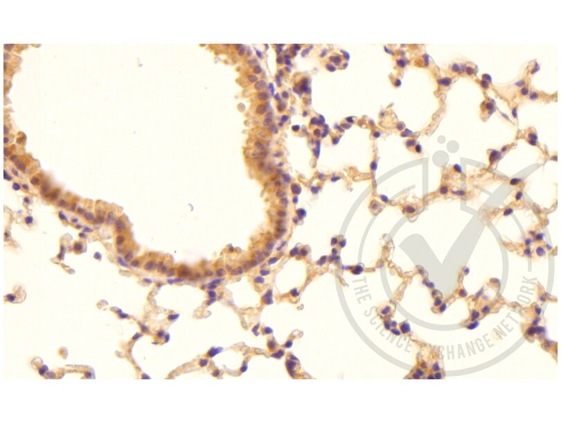
- Positive Control
- Mouse lung tissue
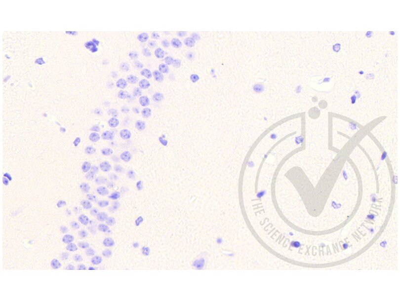
- Negative Control
- Mouse brain tissue
- Notes
- Coagulation Factor III (thromboplastin, Tissue Factor) (F3) staining is observed in positive control tissue and not in negative control tissue.
- Primary Antibody
- Antigen: Coagulation Factor III (thromboplastin, Tissue Factor) (F3)
- Catalog number: ABIN708086
- Lot number: 120224
- Secondary Antibody
- Antibody: Bond Polymer Refine Detection Kit
- Lot number: 24144
- Full Protocol
- Immunohistochemistry was performed on a Leica Bond automated immunostainer.
- Sections were deparaffinized with Novocastra Bond Dewax Solution and rehydrated into Leica Bond Wash Buffer.
- Sections were heated to 98 °C for 20 minutes in 10 mM citrate buffer pH 9.0 (ER1; Leica) for antigen retrieval.
- Sections were blocked in 3 % casein plus 0.1 % Triton-X100 for 10 minutes at room temperature.
- Sections were washed x 3 in Leica Bond Wash Buffer.
- Sections were incubated with primary antibody diluted 1:200 in Universal Antibody Dilution Buffer (Electron Microscopy Sciences, 25886-05) for 15 minutes at room temperature.
- Sections were washed x 3 in Leica Bond Wash Buffer.
- Sections were incubated with Leica Bond Polymer for 8 minutes at room temperature.
- Sections were washed x 4 in Leica Bond Wash Buffer.
- Sections were washed x 1 in Distilled Water.
- Sections were incubated with Peroxide Block (Leica) for 10 min to block endogenous peroxidase.
- Sections were washed x 4 in Leica Bond Wash Buffer.
- Sections were incubated with DAB chromogenic substrate (Leica) for 10 min at RT.
- Sections were washed x 3 in Distilled Water.
- Sections were counterstained with hematoxylin (Leica) for 2 minutes.
- Sections were washed x 1 in Distilled Water.
- Sections were washed x 1 in Leica Bond Wash Buffer.
- Sections were washed x 1 in Distilled Water.
- Sections were dehydrated, mounted and photographed under a light microscope.
- Experimental Notes
- None notes.
Validation #029589 (Immunohistochemistry)
 Validation Images
![Figure 1: Mouse lung tissue stained with anti-F3 (brown) and counterstained in blue.]() Figure 1: Mouse lung tissue stained with anti-F3 (brown) and counterstained in blue.
Figure 1: Mouse lung tissue stained with anti-F3 (brown) and counterstained in blue.
![Figure 2: Mouse brain tissue stained with anti-F3 (brown) and counterstained in blue.]() Figure 2: Mouse brain tissue stained with anti-F3 (brown) and counterstained in blue.
Figure 2: Mouse brain tissue stained with anti-F3 (brown) and counterstained in blue.
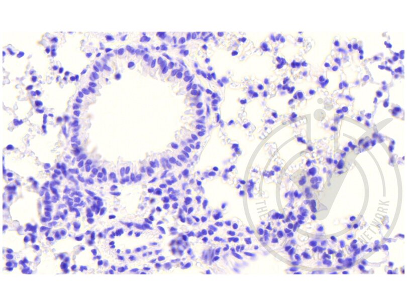
![Figure 3: Mouse lung tissue stained with isotype control antibody (brown) and counterstained in blue.]() Figure 3: Mouse lung tissue stained with isotype control antibody (brown) and counterstained in blue.
Figure 3: Mouse lung tissue stained with isotype control antibody (brown) and counterstained in blue.
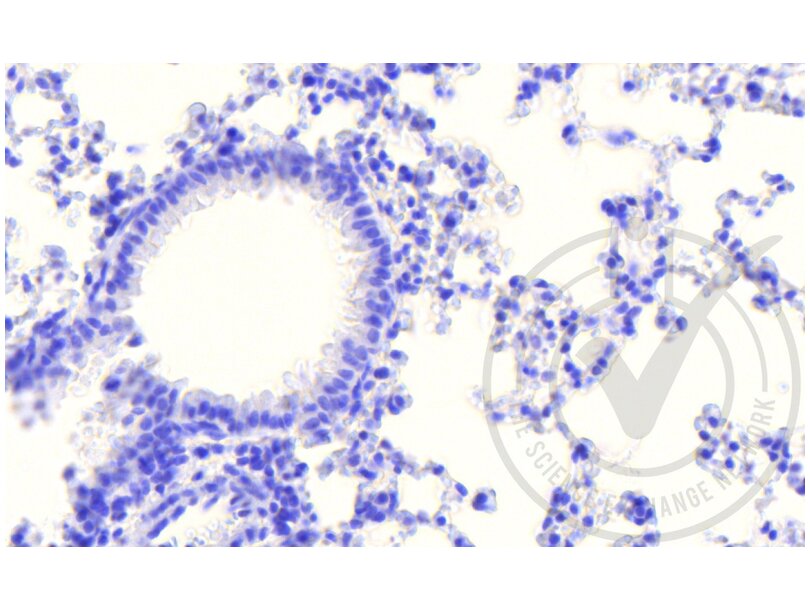
![Figure 4: Mouse lung tissue stained with secondary antibody only (brown) and counterstained in blue.]() Figure 4: Mouse lung tissue stained with secondary antibody only (brown) and counterstained in blue.
Full Methods
Figure 4: Mouse lung tissue stained with secondary antibody only (brown) and counterstained in blue.
Full Methods -
-
Format
- Liquid
-
Concentration
- 1 μg/μL
-
Buffer
- 0.01M TBS( pH 7.4) with 1 % BSA, 0.02 % Proclin300 and 50 % Glycerol.
-
Preservative
- ProClin
-
Precaution of Use
- This product contains ProClin: a POISONOUS AND HAZARDOUS SUBSTANCE, which should be handled by trained staff only.
-
Storage
- 4 °C,-20 °C
-
Storage Comment
- Shipped at 4°C. Store at -20°C for one year. Avoid repeated freeze/thaw cycles.
-
Expiry Date
- 12 months
-
-
-
: "Deconstructing Olfactory Stem Cell Trajectories at Single-Cell Resolution." in: Cell stem cell, Vol. 20, Issue 6, pp. 817-830.e8, (2017) (PubMed).
: "Histones Induce the Procoagulant Phenotype of Endothelial Cells through Tissue Factor Up-Regulation and Thrombomodulin Down-Regulation." in: PLoS ONE, Vol. 11, Issue 6, pp. e0156763, (2016) (PubMed).
: "Porcine endothelium induces DNA-histone complex formation in human whole blood: a harmful effect of histone on coagulation and endothelial activation." in: Xenotransplantation, Vol. 23, Issue 6, pp. 464-471, (2016) (PubMed).
: "Bone Marrow-Derived Mesenchymal Stem Cells Have Innate Procoagulant Activity and Cause Microvascular Obstruction Following Intracoronary Delivery: Amelioration by Antithrombin Therapy." in: Stem cells (Dayton, Ohio), (2015) (PubMed).
: "Brain-derived microparticles induce systemic coagulation in a murine model of traumatic brain injury." in: Blood, (2015) (PubMed).
: "Particulate matter phagocytosis induces tissue factor in differentiating macrophages." in: Journal of applied toxicology : JAT, (2015) (PubMed).
: "Procoagulant activity and cellular origin of microparticles in human amniotic fluid." in: Thrombosis research, Vol. 133, Issue 4, pp. 645-51, (2014) (PubMed).
-
: "Deconstructing Olfactory Stem Cell Trajectories at Single-Cell Resolution." in: Cell stem cell, Vol. 20, Issue 6, pp. 817-830.e8, (2017) (PubMed).
-
- Tissue factor (F3) (Coagulation Factor III (thromboplastin, Tissue Factor) (F3))
-
Alternative Name
- CD142
-
Background
-
Synonyms: TF, TFA, CD142, Tissue factor, Coagulation factor III, Thromboplastin, F3
Background: Initiates blood coagulation by forming a complex with circulating factor VII or VIIa. The [TF:VIIa] complex activates factors IX or X by specific limited protolysis. TF plays a role in normal hemostasis by initiating the cell-surface assembly and propagation of the coagulation protease cascade.
-
Gene ID
- 2152
-
UniProt
- P13726
-
Pathways
- Positive Regulation of Endopeptidase Activity, Smooth Muscle Cell Migration, Platelet-derived growth Factor Receptor Signaling
Target
-


