RGR antibody
 (1 reference)
 (1 validation)
Quick Overview for RGR antibody (ABIN7271760)
Target
See all RGR AntibodiesReactivity
Host
Clonality
Conjugate
Application
-
-
Purpose
- Rabbit antibody to RGR
-
Specificity
- Specific for RGR.
-
Cross-Reactivity
- Marmoset, Mouse
-
Cross-Reactivity (Details)
- Other species not yet tested.
-
Purification
- Whole serum
-
Immunogen
- A synthetic peptide from mouse retinal G protein coupled receptor (RGR) conjugated to blue carrier protein was used as the antigen.
-
-
anti-Retinal G Protein Coupled Receptor (RGR) (AA 265-291) antibody
RGR Reactivity: Human WB, IHC (p), FACS Host: Rabbit Polyclonal RB24687 unconjugated
anti-Retinal G Protein Coupled Receptor (RGR) antibodyRGR Reactivity: Human, Mouse IHC, WB, ELISA Host: Rabbit Polyclonal unconjugated
anti-Retinal G Protein Coupled Receptor (RGR) (Extracellular Domain) antibodyRGR Reactivity: Human, Monkey IHC, IHC (p) Host: Rabbit Polyclonal unconjugated
anti-Retinal G Protein Coupled Receptor (RGR) (Cytoplasmic Domain) antibodyRGR Reactivity: Human, Monkey IHC, IHC (p) Host: Rabbit Polyclonal unconjugated
anti-Retinal G Protein Coupled Receptor (RGR) (Extracellular Domain) antibodyRGR Reactivity: Human, Horse, Monkey, Xenopus laevis IHC, IHC (p) Host: Rabbit Polyclonal unconjugated
anti-Retinal G Protein Coupled Receptor (RGR) (AA 262-291), (Middle Region) antibodyRGR Reactivity: Human WB, IHC (p), FACS Host: Rabbit Polyclonal unconjugated
anti-Retinal G Protein Coupled Receptor (RGR) (AA 1-295) antibodyRGR Reactivity: Human WB, ELISA Host: Mouse Polyclonal unconjugated
anti-Retinal G Protein Coupled Receptor (RGR) (AA 152-291) antibodyRGR Reactivity: Human IHC, ELISA Host: Rabbit Polyclonal unconjugated
anti-Retinal G Protein Coupled Receptor (RGR) (AA 156-295) antibodyRGR Reactivity: Human WB Host: Rabbit Polyclonal unconjugated
anti-Retinal G Protein Coupled Receptor (RGR) antibodyRGR Reactivity: Human WB Host: Rabbit Polyclonal unconjugated
-
-
Application Notes
- IHC WB. A concentration of 10-50 μg,ml is recommended. The optimal concentration should be determined by the end user. Not yet tested in other applications.
-
Comment
-
This antibody is also available as whole serum under the product number ABIN350792. The immunogenic peptide that can be used as blocking peptide is available under the number ABIN7271761.
-
Restrictions
- For Research Use only
-
-
- by
- Palczewski Lab, Center For Translational Vision Research, UC Irvine
- No.
- #104459
- Date
- 09/21/2022
- Antigen
- RGR
- Lot Number
- Rb3134
- Method validated
- Immunohistochemistry
- Positive Control
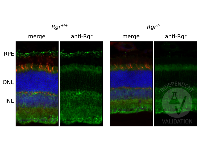
Retina cryosection from Rgr+/+ mouse, animal validated by genotyping
- Negative Control
Retina cryosection from Rgr-/- mouse, animal validated by genotyping
- Notes
Passed. Presence of specific signal in the RPE cell layer in Rgr+/+ section, and its absence in a respective Rgr-/- section was considered as indication of ABIN7271760 being immunoreactive to Rgr.
- Primary Antibody
- ABIN7271760
- Secondary Antibody
- donkey anti-rabbit AF488-conjugated antibody (Thermo Fisher Scientific, A-21206)
- Full Protocol
- Collect eyes from mice and fix with paraformaldehyde 4% (Electron Microscopy Sciences, 15710) in 1x PBS for 30 min at RT.
- Cryoprotection with sucrose series:
- Wash in 10% sucrose in 1x PBS.
- Immerse in 10% sucrose in 1x PBS for 30 min at RT.
- Wash in 20% sucrose in 1x PBS.
- Immerse in 20% sucrose in 1x PBS for 30 min RT.
- Wash in 30% sucrose in 1x PBS.
- 30% sucrose overnight at 4°C.
- Embed eyes in OCT compound (Tissue-Tek O.C.T. Compound, 4583).
- Cut retinal sections at a thickness of 12 μm on a cryostat.
- Air dry sections for 15 min at RT, store at -80°C until use.
- Sections brought to room temp, rehydrated in 1x PBS for 1h.
- Incubate sections in blocking buffer (1x PBS, 3% BSA (Sigma-Aldrich, A7030), 3% Donkey serum (Sigma-Aldrich, S30-100ML), 0.1% Triton X-100 (Sigma-Aldrich, X100-500ML)) for 1 h at RT.
- Incubate sections with primary rabbit anti-Rgr antibody (antibodies-online, ABIN7271760, lot Rb3134) diluted 1:100 in blocking buffer ON at RT. Include a no primary antibody negative controls.
- Rinse sections 3 times with 1x PBS, 0.1% Triton X-100. Keep negative controls in a separate container.
- Incubate sections with secondary donkey anti-rabbit AF488 -conjugated antibody (Thermo Fisher Scientific, A-21206) diluted 1:500 in blocking buffer for 1 h at RT.
- Rinse sections with once PBS, 0.1% Triton X-100 for 5 min at RT.
- Incubate sections in DAPI (Thermo Fisher Scientific, 62248) in 1x PBS, 0.1% Triton X-100 for 15 min.
- Rinse sections 3 times with 1x PBS, 0.1% Triton X-100 for 5 min at RT.
- Mount sections in VECTASHIELD® HardSet™ Antifade Mounting Medium (Vector Laboratories, H-1400) mounting medium.
- Acquire images with a fluorescence microscope and appropriate filter settings. For the validation purposes Keyence BZ-X800E fluorescence microscope was used with following filters: BZ-X DAPI for DAPI, BZ-X GFP for AF488, BZ-X Cy5 for Cy5. Images were taken at 100x magnification.
- Experimental Notes
The experiments involved comparison of seven different anti-mouse Rgr antisera produced in rabbit. Anti-Rgr-antiserum Rb3134-24117-WS (ABIN7271760) proved to be immunoreactive and showed much lower background level.
Rgr in mouse eye is predominantly expressed in the RPE cell layer, and to much lesser extent in the Muller glia (for details see Fig 10, PMID31694912).
To aid orientation in the tissue layers PNA (cone photoreceptor) counterstain was included in the staining (Vector Labs, PNA-Cy5 CL-1075-1) together with the secondary Ab incubation, at 1:500 diulution.
Validation #104459 (Immunohistochemistry)
 Validation Images
![Retinal sections from the Rgr+/+ and Rgr-/- animals immunostained with ABIN7271760. DAPI staining shows localization of the inner (INL) and outer (ONL) nuclear layer of the mouse retina. PNA staining was used to visualize cone inner and outer segments. RPE rests above cones, and is the site of highest Rgr expression in mouse eye. Magnification 100x.]() Retinal sections from the Rgr+/+ and Rgr-/- animals immunostained with ABIN7271760. DAPI staining shows localization of the inner (INL) and outer (ONL) nuclear layer of the mouse retina. PNA staining was used to visualize cone inner and outer segments. RPE rests above cones, and is the site of highest Rgr expression in mouse eye. Magnification 100x.
Full Methods
Retinal sections from the Rgr+/+ and Rgr-/- animals immunostained with ABIN7271760. DAPI staining shows localization of the inner (INL) and outer (ONL) nuclear layer of the mouse retina. PNA staining was used to visualize cone inner and outer segments. RPE rests above cones, and is the site of highest Rgr expression in mouse eye. Magnification 100x.
Full Methods -
-
Format
- Lyophilized
-
Reconstitution
- Reconstitute in 500 µL of sterile water. Centrifuge to remove any insoluble material.
-
Storage
- 4 °C,-20 °C
-
Storage Comment
- Maintain the lyophilised/reconstituted antibodies frozen at -20C for long term storage and refrigerated at 2-8C for a shorter term. When reconstituting glycerol (1:1) may be added for an additional stability. Avoid freeze and thaw cycles.
-
Expiry Date
- 12 months
-
-
-
: "Rapid RGR-dependent visual pigment recycling is mediated by the RPE and specialized Müller glia." in: Cell reports, Vol. 42, Issue 8, pp. 112982, (2023) (PubMed).
-
: "Rapid RGR-dependent visual pigment recycling is mediated by the RPE and specialized Müller glia." in: Cell reports, Vol. 42, Issue 8, pp. 112982, (2023) (PubMed).
-
- RGR (Retinal G Protein Coupled Receptor (RGR))
-
Alternative Name
- RGR
-
Background
- FUNCTION: Receptor for all-trans- and 11-cis-retinal. Binds preferentially to the former and may catalyze the isomerization of the chromophore by a retinochrome-like mechanism. SUBCELLULAR LOCATION: Membrane, Multi-pass membrane protein.
-
UniProt
- Q9Z2B3
Target
-


