WNT2 antibody (AA 221-320)
 (1 reference)
 (1 validation)
Quick Overview for WNT2 antibody (AA 221-320) (ABIN762896)
Target
See all WNT2 AntibodiesReactivity
Host
Clonality
Conjugate
Application
-
-
Binding Specificity
- AA 221-320
-
Cross-Reactivity
- Human, Mouse
-
Purification
- Purified by Protein A.
-
Immunogen
- KLH conjugated synthetic peptide derived from human WNT2
-
Isotype
- IgG
-
-
anti-Wingless-Type MMTV Integration Site Family Member 2 (WNT2) (AA 26-360) antibody
WNT2 Reactivity: Human WB, IHC, ELISA Host: Rabbit Polyclonal unconjugated
anti-Wingless-Type MMTV Integration Site Family Member 2 (WNT2) antibodyWNT2 Reactivity: Human, Mouse IHC, ELISA Host: Rabbit Polyclonal unconjugated
anti-Wingless-Type MMTV Integration Site Family Member 2 (WNT2) (AA 26-360) antibodyWNT2 Reactivity: Human WB, IHC, IP, ICC Host: Rabbit Polyclonal unconjugated
anti-Wingless-Type MMTV Integration Site Family Member 2 (WNT2) (AA 254-287) antibodyWNT2 Reactivity: Human WB Host: Rabbit Polyclonal RB49732 unconjugated
anti-Wingless-Type MMTV Integration Site Family Member 2 (WNT2) (Internal Region) antibodyWNT2 Reactivity: Human, Monkey IHC, IHC (p) Host: Rabbit Polyclonal unconjugated
anti-Wingless-Type MMTV Integration Site Family Member 2 (WNT2) (AA 143-192) antibodyWNT2 Reactivity: Human, Mouse, Rat, Cow, Dog, Guinea Pig, Horse, Rabbit, Sheep, Zebrafish (Danio rerio), Monkey, Bat, Pig WB Host: Rabbit Polyclonal unconjugated
-
-
Application Notes
-
WB 1:300-5000
ELISA 1:500-1000
IHC-P 1:200-400
IHC-F 1:100-500
IF(IHC-P) 1:50-200
IF(IHC-F) 1:50-200
IF(ICC) 1:50-200 -
Restrictions
- For Research Use only
-
-
- by
- Histopathology and Tissue Shared Resource, Georgetown Lombardi Comprehensive Cancer Center
- No.
- #029629
- Date
- 03/16/2014
- Antigen
- Lot Number
- 131105
- Method validated
- Immunohistochemistry
- Positive Control
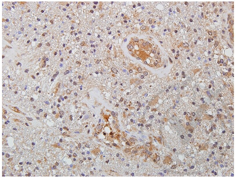
- Human glioma tissue
- Negative Control
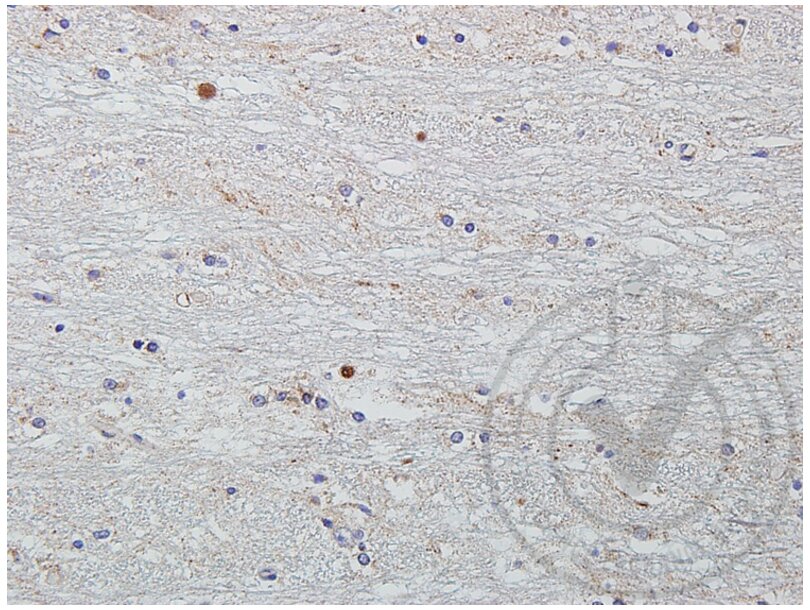
- Human brain
- Notes
- Signal was detected in positive control tissue and not in negative control tissue.
- Primary Antibody
- Antigen: Wingless-Type MMTV Integration Site Family Member 2 (WNT2) (N-Term)
- Catalog number: ABIN762896
- Lot number: 131105
- Secondary Antibody
- Antibody: Envision Plus Horse Radish Peroxidase conjugated anti-rabbit antibody
- Batch number: 10082183
- Full Protocol
- Immunohistochemistry was performed by hand.
- Sections were de-paraffinized on a Leica Autostainer in Xylenes (1X 5 min, 2X 2 min), and rehydrated through Ethanols (2X 100% for 5 min each, 95% 2X 2 min each, 80%, 70%) to running tap water. - Sections were heated to 98°C for 20 min in 10 mM Sodium Citrate buffer pH 6.0 for antigen retrieval, then moved to RT in the same buffer for 20 min.
- Sections were rinsed in de-ionized water for 5 min at RT.
- Sections were blocked in Hydrogen Peroxide (Fisher, H325-500) for 30 min at RT.
- Sections were rinsed in Tris Buffered Saline with 0.5% Tween-20 (TBST) 2 times for 5 min at RT.
- Sections were blocked in 5% Normal Goat Serum for 2 h at RT.
- Sections were incubated with primary antibody diluted 1:250 in TBST overnight at 4°C.
- Sections were rinsed in TBST for 5 min 2X at RT.
- Sections were incubated with Envision Plus anti-Rabbit-Horse Radish Peroxidase conjugated Polymer for 2 h at RT.
- Sections were rinsed in TBST for 5 min 2X at RT.
- Sections were incubated with DAB chromogenic substrate (DAKO, K348) for 10 min at RT.
- Sections were washed x 1 in Distilled Water.
- Sections were counterstained with 1:9 dilution of Harris Hematoxylin (Fisher, SH30-500D) for 2 min.
- Sections were washed x 1 in Distilled Water.
- Sections were blued in Ammonium Hydroxide for 1 min.
- Sections were washed x 1 in Distilled Water.
- Sections were dehydrated through graded alcohols, mounted in Acrymount and photographed on an Olympus DX61 microscope with DP70 camera using DP Controller and DP Manager Software.
- Experimental Notes
- The company recommends between 1:100 – 1:500 dilution factor; in our hands, 1:250 was overstained and we would recommend using a lower concentration of the antibody.
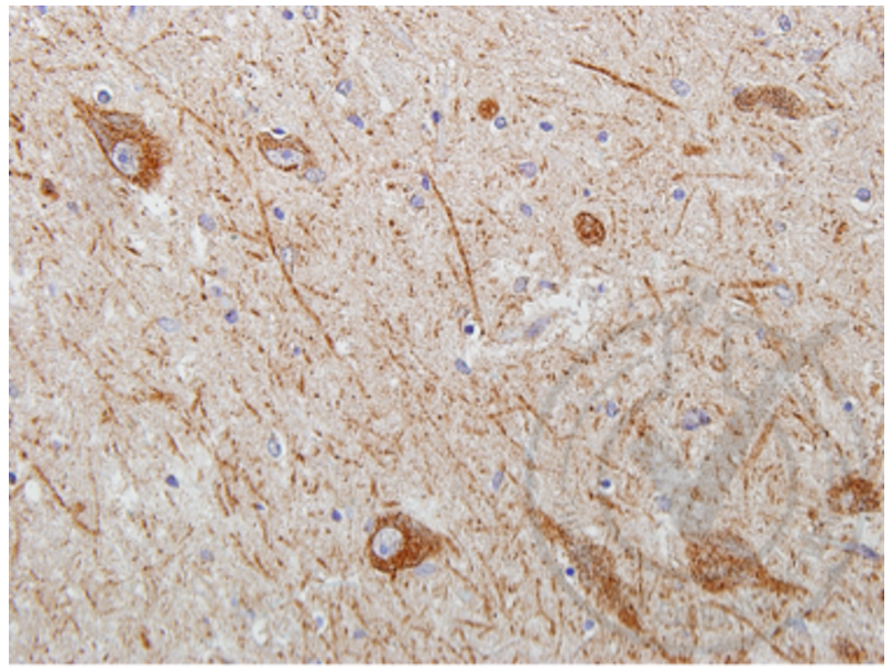
- The “normal” brain section we used proved to be from an individual who had morphological features of Parkinson’s Disease. The area shown in Figure 2 is from the morphologically normal part of the brain. The area with Parkinson’s morphology reacted strongly with the anti-wnt2 antibody (Figure 5).
Validation #029629 (Immunohistochemistry)
 Validation Images
![Figure 1: Human brain glioma tissue stained with anti-Wnt2 (brown) and counterstained with hematoxylin (blue).]() Figure 1: Human brain glioma tissue stained with anti-Wnt2 (brown) and counterstained with hematoxylin (blue).
Figure 1: Human brain glioma tissue stained with anti-Wnt2 (brown) and counterstained with hematoxylin (blue).
![Figure 2: Human normal brain tissue stained with anti-Wnt2 (brown) and counterstained with hematoxylin (blue).]() Figure 2: Human normal brain tissue stained with anti-Wnt2 (brown) and counterstained with hematoxylin (blue).
Figure 2: Human normal brain tissue stained with anti-Wnt2 (brown) and counterstained with hematoxylin (blue).
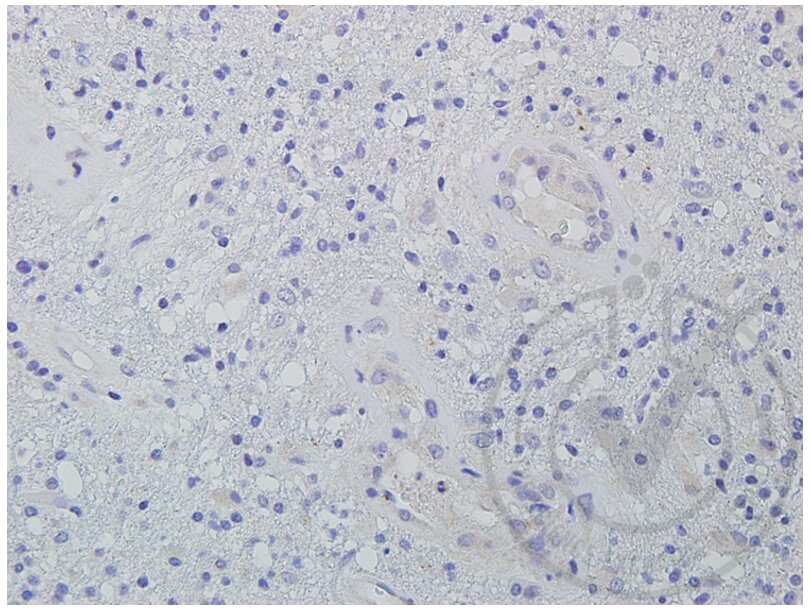
![Figure 3: Human brain glioma tissue stained with isotype control antibody (brown) and counterstained with hematoxylin (blue).]() Figure 3: Human brain glioma tissue stained with isotype control antibody (brown) and counterstained with hematoxylin (blue).
Figure 3: Human brain glioma tissue stained with isotype control antibody (brown) and counterstained with hematoxylin (blue).
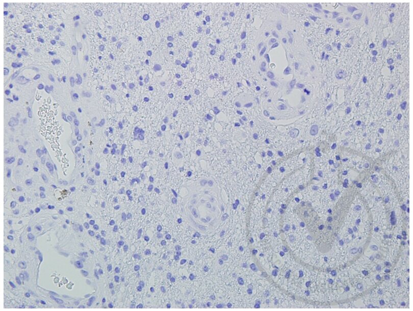
![Figure 4: Human brain glioma tissue stained with secondary only (brown) and counterstained with hematoxylin (blue).]() Figure 4: Human brain glioma tissue stained with secondary only (brown) and counterstained with hematoxylin (blue).
Figure 4: Human brain glioma tissue stained with secondary only (brown) and counterstained with hematoxylin (blue).
![Figure 5: Human brain tissue with Parkinson's morphology stained with anti-Wnt2 (brown) and counterstained with hematoxylin (blue).]() Figure 5: Human brain tissue with Parkinson's morphology stained with anti-Wnt2 (brown) and counterstained with hematoxylin (blue).
Full Methods
Figure 5: Human brain tissue with Parkinson's morphology stained with anti-Wnt2 (brown) and counterstained with hematoxylin (blue).
Full Methods -
-
Format
- Liquid
-
Concentration
- 1 μg/μL
-
Buffer
- 0.01M TBS( pH 7.4) with 1 % BSA, 0.02 % Proclin300 and 50 % Glycerol.
-
Preservative
- ProClin
-
Precaution of Use
- This product contains ProClin: a POISONOUS AND HAZARDOUS SUBSTANCE, which should be handled by trained staff only.
-
Storage
- 4 °C,-20 °C
-
Storage Comment
- Shipped at 4°C. Store at -20°C for one year. Avoid repeated freeze/thaw cycles.
-
Expiry Date
- 12 months
-
-
-
: "Macrophages mediate a switch between canonical and non-canonical Wnt pathways in canine mammary tumors." in: PLoS ONE, Vol. 9, Issue 1, pp. e83995, (2014) (PubMed).
-
: "Macrophages mediate a switch between canonical and non-canonical Wnt pathways in canine mammary tumors." in: PLoS ONE, Vol. 9, Issue 1, pp. e83995, (2014) (PubMed).
-
- WNT2 (Wingless-Type MMTV Integration Site Family Member 2 (WNT2))
-
Alternative Name
- WNT2
-
Background
-
Synonyms: IRP, INT1L1, Protein Wnt-2, Int-1-like protein 1, Int-1-related protein, WNT2
Background: Ligand for members of the frizzled family of seven transmembrane receptors. Probable developmental protein. May be a signaling molecule which affects the development of discrete regions of tissues. Is likely to signal over only few cell diameters.
-
Gene ID
- 7472
-
UniProt
- P09544
-
Pathways
- WNT Signaling
Target
-


