CCDC42 antibody (AA 221-312)
 (2 validations)
Quick Overview for CCDC42 antibody (AA 221-312) (ABIN872760)
Target
See all CCDC42 AntibodiesReactivity
Host
Clonality
Conjugate
Application
-
-
Binding Specificity
- AA 221-312
-
Cross-Reactivity
- Mouse, Rat
-
Predicted Reactivity
- Human,Dog,Cow,Sheep,Pig,Horse,Rabbit
-
Purification
- Purified by Protein A.
-
Immunogen
- KLH conjugated synthetic peptide derived from human CCDC42
-
Isotype
- IgG
-
-
anti-Coiled-Coil Domain Containing 42 (CCDC42) (Middle Region) antibody
CCDC42 Reactivity: Human, Mouse, Rat, Cow, Dog, Guinea Pig, Horse, Rabbit WB Host: Rabbit Polyclonal unconjugated
anti-Coiled-Coil Domain Containing 42 (CCDC42) (AA 121-150) antibodyCCDC42 Reactivity: Human, Mouse WB, IHC (p) Host: Rabbit Polyclonal RB25993 unconjugated
anti-Coiled-Coil Domain Containing 42 (CCDC42) (AA 1-242) antibodyCCDC42 Reactivity: Human WB, ELISA, IHC Host: Rabbit Polyclonal unconjugated
anti-Coiled-Coil Domain Containing 42 (CCDC42) (AA 161-210) antibodyCCDC42 Reactivity: Human, Rat WB Host: Rabbit Polyclonal unconjugated
-
-
Application Notes
-
WB 1:300-5000
ELISA 1:500-1000
IHC-P 1:200-400
IHC-F 1:100-500
IF(IHC-P) 1:50-200
IF(IHC-F) 1:50-200
IF(ICC) 1:50-200 -
Restrictions
- For Research Use only
-
-
- by
- Developmental Biology, Johann-Friedrich-Blumenbach-Institute for Zoology and Anthropology, Georg-August-University of Göttingen
- No.
- #103396
- Date
- 10/12/2018
- Antigen
- CCDC42
- Lot Number
- AH09037940
- Method validated
- Immunocytochemistry
- Positive Control
- NIH/3T3 cells transfected with Ccdc42-mCherry expression vector
- Negative Control
- NIH/3T3 cell lysates
- Notes
ABIN872760 specifically detects exogenously expressed Ccdc42-Cherry but shows also unspecific background staining.
- Primary Antibody
- ABIN872760
- Secondary Antibody
- goat anti-rabbit 488 (MoBiotec MFP-A1008, lot 2806F058)
- Full Protocol
- Grow NIH/3T3 cells (ATCC, CRL-1658) in DMEM+GlutaMAX (Gibco, 10567-014, lot 1922818) supplemented with fetal bovine serum (Gibco, 10500-064, lot 08FO477K) and Pen/Strep (Gibco 15140-122, lot 1924797), at 37°C and 5% CO2 to 70% confluency.
- Transfect cells with mCherry-tagged Ccdc42 (Ccdc42-Cherry) expression vector using EndofectinMax (GeneCopoeia) following the manufacturer´s instructions.
- Grow cells for 24h.
- Fix cells in methanol for 10min at -20°C followed by incubation in 0.3% Triton X-100 for 10min at 4°C.
- Block cells in PBS containing 1% bovine serum albumin and 0.5% Tween-20 (PBT) for 1h at RT.
- Incubate cells with primary rabbit anti-CCDC42 antibody (antibodies-online, ABIN872760, lot AH09037940) diluted 1:100 in PBS for 1h at 37°C.
- Wash cells 3x 5min with 1x PBS.
- Incubate cells with secondary goat anti-rabbit 488 antibody (MoBiotec MFP-A1008, lot 2806F058) diluted 1:300 in PBS for 1h at 37°C.
- DNA counterstained with DAPI (4’,6-Diamidino-2-phenylindole; Sigma D-9542).
- Image acquisition on Zeiss LSM 510 confocal microscope and processing using Adobe Photoshop 5.0.
- Experimental Notes
Validation #103396 (Immunocytochemistry)
 Validation Images
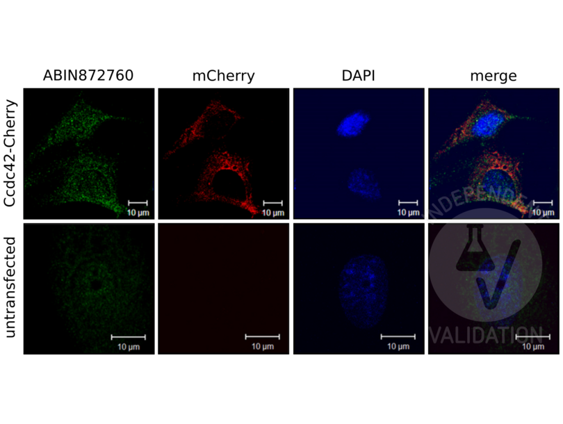
![Immunocytochemistry detection of NIH/3T3 cells expressing mCherry-labeled Ccdc42 (Ccdc42-Cherry) using ABIN872760 (green) or an mCherry antibody (red). Untransfected NIH/3T3 cells served as negative control. The pictures on the right show the red and green channels merged with DAPI counterstain (blue).]() Immunocytochemistry detection of NIH/3T3 cells expressing mCherry-labeled Ccdc42 (Ccdc42-Cherry) using ABIN872760 (green) or an mCherry antibody (red). Untransfected NIH/3T3 cells served as negative control. The pictures on the right show the red and green channels merged with DAPI counterstain (blue).
Full Methods
Immunocytochemistry detection of NIH/3T3 cells expressing mCherry-labeled Ccdc42 (Ccdc42-Cherry) using ABIN872760 (green) or an mCherry antibody (red). Untransfected NIH/3T3 cells served as negative control. The pictures on the right show the red and green channels merged with DAPI counterstain (blue).
Full Methods -
- by
- Developmental Biology, Johann-Friedrich-Blumenbach-Institute for Zoology and Anthropology, Georg-August-University of Göttingen
- No.
- #103397
- Date
- 10/12/2018
- Antigen
- CCDC42
- Lot Number
- AH09037940
- Method validated
- Western Blotting
- Positive Control
- Lysate from HEK293 cells transfected with mCherry tagged Ccdc42 expression vector
- Negative Control
- Lysate from untransfected HEK293 cells
- Notes
Passed. ABIN872760 specifically detects Ccdc42 by immunoblotting.
- Primary Antibody
- ABIN872760
- Secondary Antibody
- goat anti-rabbit IgG (H+L), HRP-conjugated (Jackson Immuno Research, 111-035-003, lot 123450)
- Full Protocol
- Grow HEK293 (ATCC) cells in DMEM+GlutaMAX (Gibco, 10567-014, lot 1922818) supplemented with fetal bovine serum (Gibco, 10500-064, lot 08FO477K) and Pen/Strep (Gibco, 15140-122, lot 1924797), at 37°C and 5% CO2 to 70% confluency.
- Transfect cells with mCherry tagged Ccdc42 (Ccdc42-Cherry) expression vector using EndofectinMax (GeneCopoeia) following the manufacturer´s instructions.
- Grow cells for 24h.
- Lyse cells in SDS-sample buffer and denature total cellular lysates for 5min at 95°C.
- Separate up to 40µl of protein lysate on a denaturing 10% SDS polyacrylamide gel (Laemmli (1970)).
- Transfer proteins onto 0.2µm Protran membrane (GE Healthcare, 10600004, A10043108) with a Western blotting system for 1h at 400A (Towbin et al. (1979)).
- Block the membrane in TBST (50mM Tris-HCl pH7.4, 150mM NaCl, 0.2% Tween 20) containing 5% dry milk (blocking solution) for 1h at RT.
- Incubate membrane with primary
- rabbit anti-Ccdc42 antibody (antibodies-online, ABIN872760, lot AH09037940) diluted 1:500 in blocking solution ON at 4°C or
- rat anti-RFP antibody (antibodies-online, ABIN334653, lot 090428) diluted 1:500 in blocking solution ON at 4°C.
- Wash membrane for 6x 5min with TBST at RT.
- Incubate membrane with secondary
- goat anti-rabbit IgG (H+L), HRP-conjugated antibody (Jackson Immuno Research, 111-035-003, lot 123450) diluted 1:2000 in blocking solution for 45min at RT or
- rabbit anti-rat IgG-HRPconjugated antibody (Jackson ImmunoResearch Lab. Inc., 312-035-003, lot 34336) diluted 1:2000 in blocking solution for 45min at RT.
- Wash membranes for 45 min at RT with TBST.
- Reveal protein bands using Clarity Max Western ECL substrate (Bio-Rad, 1705062) and capture images via Chemidoc Imaging System (Bio-Rad).
- Experimental Notes
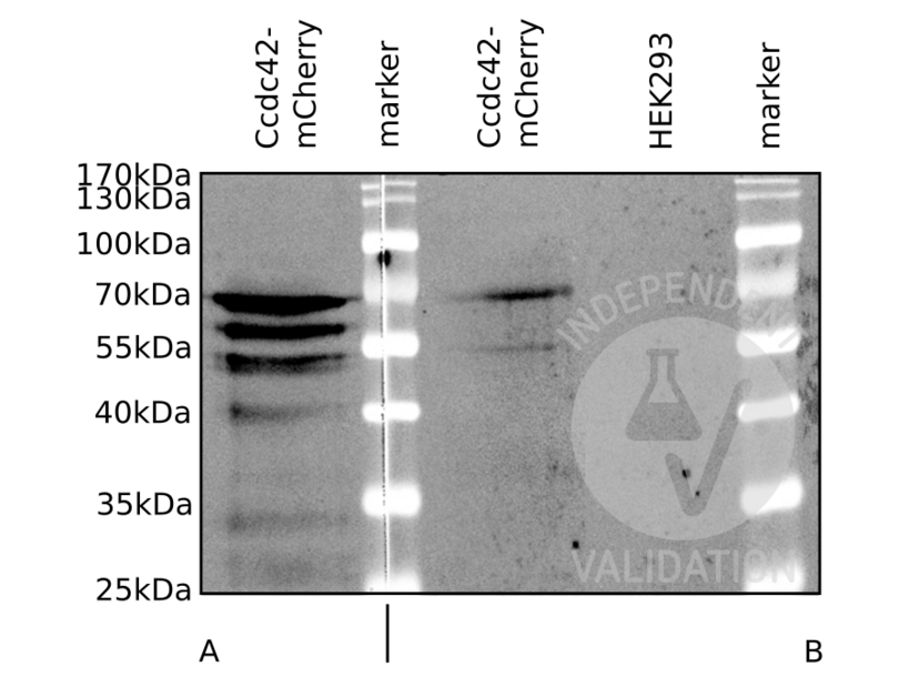
Expected molecular mass of Ccdc42-tagged Cherry is approximately 68kDa. ABIN872760 detects a protein in the expected molecular mass range. The anti-RedFP antibody detects a protein with a similar mass in addition to proteins with lower molecular masses, which are most probably degradation products.
Validation #103397 (Western Blotting)
 Validation Images
![Western blot analysis of lysates from HEK293 cells expressing mCherry tagged Ccdc42 (Ccdc42-mCherry) or untransfected HEK293 cells (HEK293). The membrane was incubated with a primary RFP antibody (A) or ABIN872760 (B).]() Western blot analysis of lysates from HEK293 cells expressing mCherry tagged Ccdc42 (Ccdc42-mCherry) or untransfected HEK293 cells (HEK293). The membrane was incubated with a primary RFP antibody (A) or ABIN872760 (B).
Full Methods
Western blot analysis of lysates from HEK293 cells expressing mCherry tagged Ccdc42 (Ccdc42-mCherry) or untransfected HEK293 cells (HEK293). The membrane was incubated with a primary RFP antibody (A) or ABIN872760 (B).
Full Methods -
-
Format
- Liquid
-
Concentration
- 1 μg/μL
-
Buffer
- 0.01M TBS( pH 7.4) with 1 % BSA, 0.02 % Proclin300 and 50 % Glycerol.
-
Preservative
- ProClin
-
Precaution of Use
- This product contains ProClin: a POISONOUS AND HAZARDOUS SUBSTANCE, which should be handled by trained staff only.
-
Storage
- 4 °C,-20 °C
-
Storage Comment
- Shipped at 4°C. Store at -20°C for one year. Avoid repeated freeze/thaw cycles.
-
Expiry Date
- 12 months
-
-
- CCDC42 (Coiled-Coil Domain Containing 42 (CCDC42))
-
Alternative Name
- CCDC42
-
Background
-
Synonyms: CCDC42A, Coiled coil domain containing 42, Coiled coil domain containing protein 42A, FLJ32734, OTTMUSP00000006142, RGD1565925, RP23-400O5.1, CCD42_HUMAN.
Background: The coiled-coil domain is a structural motif found in proteins that are involved in a diverse array of biological functions such as the regulation of gene expression, cell division, membrane fusion and drug extrusion and delivery. CCDC42 (coiled-coil domain containing 42) is a 316 amino acid protein encoded by a gene that maps to human chromosome 17p13.1. Encoding over 1,200 genes, chromosome 17 comprises over 2.5 % of the human genome. Two key tumor suppressor genes are associated with chromosome 17, namely, p53 and BRCA1. Tumor suppressor p53 is necessary for maintenance of cellular genetic integrity by moderating cell fate through DNA repair versus cell death. Malfunction or loss of p53 expression is associated with malignant cell growth and Li-Fraumeni syndrome.
-
Gene ID
- 146849
Target
-


