CD200R1 antibody (C-Term)
 (1 reference)
 (1 validation)
-
- Target See all CD200R1 Antibodies
- CD200R1 (CD200 Receptor 1 (CD200R1))
-
Binding Specificity
- AA 275-303, C-Term
-
Reactivity
- Human
-
Host
- Rabbit
-
Clonality
- Polyclonal
-
Conjugate
- This CD200R1 antibody is un-conjugated
-
Application
- Western Blotting (WB), Immunohistochemistry (IHC), Immunohistochemistry (Paraffin-embedded Sections) (IHC (p))
- Predicted Reactivity
- Bovine
- Purification
- This antibody is purified through a protein A column, followed by peptide affinity purification.
- Immunogen
- This CD200R1 antibody is generated from rabbits immunized with a KLH conjugated synthetic peptide between 275-303 amino acids from the C-terminal region of human CD200R1.
- Clone
- RB35254
- Isotype
- Ig Fraction
-
anti-CD200 Receptor 1 (CD200R1) (AA 1-348) antibody
CD200R1 Reactivity: Human WB, IP Host: Rabbit Polyclonal unconjugated
anti-CD200 Receptor 1 (CD200R1) (AA 391-405), (C-Term), (Intracellular) antibodyCD200R1 Reactivity: Human, Mouse, Rat WB, IHC, ICC, IF Host: Rabbit Polyclonal unconjugated
anti-CD200 Receptor 1 (CD200R1) antibodyCD200R1 Reactivity: Human FACS, ICC Host: Mouse Monoclonal OX-108 unconjugated
anti-CD200 Receptor 1 (CD200R1) (AA 201-300) antibodyCD200R1 Reactivity: Human WB, ELISA, IF (cc), IF (p), ICC, IHC (p), IHC (fro) Host: Rabbit Polyclonal unconjugated
anti-CD200 Receptor 1 (CD200R1) antibody (PE)CD200R1 Reactivity: Human FACS Host: Mouse Monoclonal OX-108 PE
anti-CD200 Receptor 1 (CD200R1) (AA 91-140) antibodyCD200R1 Reactivity: Human WB Host: Rabbit Polyclonal unconjugated
anti-CD200 Receptor 1 (CD200R1) (C-Term) antibodyCD200R1 Reactivity: Human WB, ELISA, ICC, IF Host: Rabbit Polyclonal unconjugated
anti-CD200 Receptor 1 (CD200R1) antibodyCD200R1 Reactivity: Human FACS Host: Rabbit Chimeric OX108 unconjugated Recombinant Antibody
anti-CD200 Receptor 1 (CD200R1) (AA 249-348) antibodyCD200R1 Reactivity: Human WB Host: Rabbit Polyclonal unconjugated
anti-CD200 Receptor 1 (CD200R1) (AA 2-14) antibodyCD200R1 Reactivity: Human, Pig, Cow, Dog, Horse, Monkey WB, ELISA Host: Goat Polyclonal unconjugated
-
- Application Notes
- WB: 1:1000IHC: 1:100
- Restrictions
- For Research Use only
-
- by
- David A. Clark, Department of Medicine and Dept. Pathology and Molecular Medicine, McMaster University
- No.
- #104430
- Date
- 04/04/2022
- Antigen
- CD200R1
- Lot Number
- SA110916AG
- Method validated
- Immunohistochemistry
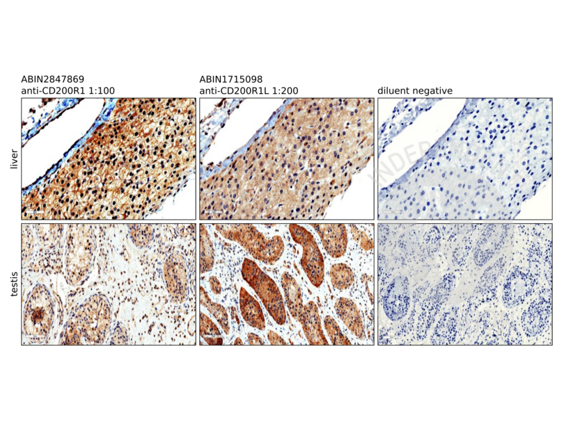
- Positive Control
Human liver (biopsy of normal liver tissue in a 3-year-old) and testis tissue (normal testis tissue from a pubertal testis surgically removed due to trauma) stained with anti-CD200R1 ABIN2847869.
- Negative Control
anti-CD200R1L antibody ABIN1715098
antibody diluent only
- Notes
Passed. The CD200R1 antibody ABIN2847869 specifically labels CD200R1 in human liver and testis in IHC-P.
- Primary Antibody
- ABIN2847869
- Secondary Antibody
- Bond Polymer Refine Detection kit (Leica, DS9800, lot 49232)
- Full Protocol
- Cut paraffin blocks with a Leica CM2255 Microtome into 4 µm sections.
- Affix sections to positively charged slides and air dry ON at RT.
- Dewax the slides and hydrate on the automated Leica BOND Rx automated slide stainer.
- Antigen retrieval on the Leica BOND Rx automated slide stainer using epitope retrieval buffer 2 (ER2, Leica, AR9640).
- Stain slides with primary
- rabbit anti- CD200R1 (AA 45-95) antibody (antibodies-online, ABIN2847869, lot SA110916AG) or
- rabbit anti-CD200 Receptor 1-Like (CD200R1L) (AA 150-200) antibody (antibodies-online, ABIN1715098),
- diluted 1:100 or 1:200 in Power Vision IHC Super Blocker (Leica, PV6122). The staining protocol incorporates a modified Leica standard protocol IHC-F (which omits the post-primary step) and uses the standard times outlined in the machine protocol.
- Stain sections with Bond Polymer Refine Detection kit (Leica, DS9800, lot 49232) containing peroxidase block, post primary antibody, polymer as well as DAB chromogen and hematoxylin counterstain for times outlined in the standard protocol IHC-F.
- Remove slides from the Leica Bond Rx and then dehydrate in ethanol and clear in xylene.
- Mount slides mounted in Fisher Chemical Permount Mounting Medium (Fisher Scientific, SP15-500, lot 162767).
- Once the slide-coverslip edges are dry, scan the slides using Leica Imagescope (40x) and photograph into jpg files at 200X and 400X.
- Quantify staining of 200X jpg images using Image J and the hand drawing tool allowing individual testicular tubules to be assessed, and similarly areas of liver tissue free of central veins and portal connective tissue. Compute the mean value and sem of staining with antibody versus diluent control negative using GraphPad Prism software (San Diego, CA, USA).
- Experimental Notes
Rationale for choice of positive control tissue: CD200R1 and CD200R1L on the NCBI website (NM_138806 and NM_001008784.2) are based on data from Fagerberg L et al. (2014) Mol Cell Proteomics. PMID 24309898. Analysis of the data was used to provide a quantification score (FPKM) reflecting gene expression for 27 tissues. Of particular interest are the results for liver and testis in supplementary data set 1. Testis was the only tissue with CD200R1L > CD200R1.
CD200R1 expression (FPKM) liver: 1.32
CD200R1 expression (FPKM) testis: 1.22
CD200R1L expression (FPKM) liver: 0.00
CD200R1L expression (FPKM) testis: 2.19
A CD200R1L expression value of zero strongly suggests that CD200R1L protein would not likely be found in liver tissue, but in testis tissue, CD200R1 staining should be less than CD200R1L staining.
One can test the specificity of the antibodies to CD200R1 and CD200R1L by comparing IHC staining intensity to the result predicted from the list above.
A 1:100 dilution of anti-CD200R1 antibody ABIN2847869 was superior to staining with a 1:200 dilution whereas a 1:200 dilution of anti-CD200R1L antibody ABIN1715098 was optimal based on previous analysis.
Although liver staining with anti-CD200R1L with antibody ABIN1715098 was slightly less intense than testis staining (P = 0.033 by Student’s t test), based on the pre-existing molecular analysis, the important point is that minimal or no staining of liver should have occurred. ABIN1715098 is an antigen-affinity-purified rabbit polyclonal antibodies to CD200R1L protein (Q6Q8B3). However, in Western blots, ABIN1715098 and similarly generated antibodies to Q6Q8B3 show 2 bands, one at about 29 kD compatible with CD200R1, and 1 at 37 kD, compatible with CD200R1. Q6Q8B3-1 expresses a 41 AA sequence (AA 56-101) and 21 AA sequence (AA 135-155) present in CD200R1 protein, so some of the antibodies generated against Q6Q8B3-1 should bind to CD200R1 (Q8TD46). ABIN2847869 binds to a 29 AA sequence in the intracytoplasmic tail (C region) of human CD200R1 which is not present in CD200R1L.
Therefore, ABIN1715098 staining is most likely due to binding to CD200R1. The previous published suggestion that ABIN1715098 is anti-CD200R is validated (Clark DA et al. (2018) J Reprod Immunol. PMID 29571945).
There can be many reasons why staining with the two antibodies was not identical. In the table shown in the figures, CD200R1/CD200R1L = 1.27, and for other tissues, one could normalize by multiplying CD200R1L by 1.27 to obtain a potentially better estimate.
For testis, anti-CD200R1L staining was greater than anti-CD200R1 staining, and the fraction CD200R1/CD200R1L = 0.315 (0.248 with normalization of CD200R1L) and given that protein production by a cell may not correlate exactly with the mRNA value for that protein (Fagerberg L et al., ibid), the IHC result is consistent with the molecular predicted value of 0.557 above. We do not know exactly how Fagerberg et al. prepared their tissues for analysis, and for testis tissue sections, we confined our attention to seminiferous tubules and ignored CD200R1-positive macrophages in testicular stroma.
Validation #104430 (Immunohistochemistry)
 Validation Images
![IHC staining of liver and testis tissue with CD200R1 antibody ABIN2847869. For liver, the 400X magnification shows labeling of hepatocyte membranes and no labelling of the wall of the central vein. Also note variation in staining intensity. The photomicrograph of the testis is shown at a lower magnification (200X) in order to show heterogeneity of seminiferous tubule staining intensity. V, central vein wall; L, liver cells; S, testicular stroma; ST, seminiferous tubule.]() IHC staining of liver and testis tissue with CD200R1 antibody ABIN2847869. For liver, the 400X magnification shows labeling of hepatocyte membranes and no labelling of the wall of the central vein. Also note variation in staining intensity. The photomicrograph of the testis is shown at a lower magnification (200X) in order to show heterogeneity of seminiferous tubule staining intensity. V, central vein wall; L, liver cells; S, testicular stroma; ST, seminiferous tubule.
IHC staining of liver and testis tissue with CD200R1 antibody ABIN2847869. For liver, the 400X magnification shows labeling of hepatocyte membranes and no labelling of the wall of the central vein. Also note variation in staining intensity. The photomicrograph of the testis is shown at a lower magnification (200X) in order to show heterogeneity of seminiferous tubule staining intensity. V, central vein wall; L, liver cells; S, testicular stroma; ST, seminiferous tubule.
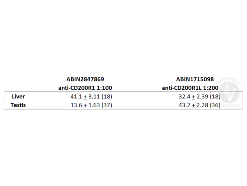
![Summary of staining intensity with antibody compared to diluent control. Parentheses show number of measurements.]() Summary of staining intensity with antibody compared to diluent control. Parentheses show number of measurements.
Full Methods
Summary of staining intensity with antibody compared to diluent control. Parentheses show number of measurements.
Full Methods -
- Format
- Liquid
- Buffer
- Purified polyclonal antibody supplied in PBS with 0.09 % (W/V) sodium azide.
- Preservative
- Sodium azide
- Precaution of Use
- This product contains Sodium azide: a POISONOUS AND HAZARDOUS SUBSTANCE which should be handled by trained staff only.
- Storage
- 4 °C,-20 °C
- Storage Comment
- CD200R1 Antibody (C-term) can be refrigerated at 2-8 °C for up to 2 weeks. For long term storage store at -20 °C in small aliquots to prevent freeze-thaw cycles.
- Expiry Date
- 6 months
-
-
: "Circulating CD200 is increased in the secretory phase of women with endometriosis as is endometrial mRNA, and endometrial stromal cell CD200R1 is increased in spite of reduced mRNA." in: American journal of reproductive immunology (New York, N.Y. : 1989), Vol. 89, Issue 1, pp. e13655, (2022) (PubMed).
-
- Target
- CD200R1 (CD200 Receptor 1 (CD200R1))
- Alternative Name
- CD200R1 (CD200R1 Products)
- Synonyms
- CD200R antibody, HCRTR2 antibody, MOX2R antibody, OX2R antibody, Mox2r antibody, CD200R1 antibody, CD200R2 antibody, CD200R-B1V1 antibody, CD200R1A antibody, CD200 receptor 1 antibody, cell surface glycoprotein CD200 receptor 1 antibody, CD200 receptor 1-like antibody, CD200R1 antibody, Cd200r1 antibody, LOC487977 antibody, CD200R1L antibody
- Background
- This gene encodes a receptor for the OX-2 membrane glycoprotein. Both the receptor and substrate are cell surface glycoproteins containing two immunoglobulin-like domains. This receptor is restricted to the surfaces of myeloid lineage cells and the receptor-substrate interaction may function as a myeloid downregulatory signal. Mouse studies of a related gene suggest that this interaction may control myeloid function in a tissue-specific manner. Alternative splicing of this gene results in multiple transcript variants.
- Molecular Weight
- 36620
- Gene ID
- 131450
- NCBI Accession
- NP_620161, NP_620385, NP_620386, NP_740750
- UniProt
- Q8TD46
-


