Metabotropic Glutamate Receptor 6 antibody (AA 828-877)


 (2 validations)
Quick Overview for Metabotropic Glutamate Receptor 6 antibody (AA 828-877) (ABIN1533291)
Target
See all Metabotropic Glutamate Receptor 6 (GRM6) AntibodiesReactivity
Host
Clonality
Conjugate
Application
-
-
Binding Specificity
- AA 828-877
-
Purpose
- mGluR-6 Antibody
-
Specificity
- MGluR-6 Polyclonal Antibody detects endogenous levels of mGluR-6 protein.
-
Purification
- The antibody was affinity-purified from rabbit antiserum by affinity-chromatography using epitope-specific immunogen.
-
Purity
- > 95 %
-
Immunogen
- The antiserum was produced against synthesized peptide derived from human mGluR6. AA range:828-877
-
Isotype
- IgG
-
-
anti-Glutamate Receptor, Metabotropic 6 (GRM6) (C-Term) antibody
GRM6 Reactivity: Human, Rat, Mouse WB, IF Host: Rabbit Polyclonal unconjugated
anti-Glutamate Receptor, Metabotropic 6 (GRM6) (AA 480-585) antibodyGRM6 Reactivity: Human ELISA, FACS Host: Mouse Monoclonal 7D9C9 unconjugated
anti-Glutamate Receptor, Metabotropic 6 (GRM6) antibodyGRM6 Reactivity: Human WB, IHC Host: Rabbit Polyclonal unconjugated
anti-Glutamate Receptor, Metabotropic 6 (GRM6) (N-Term) antibodyGRM6 Reactivity: Human, Monkey IHC, IHC (p) Host: Rabbit Polyclonal unconjugated
anti-Glutamate Receptor, Metabotropic 6 (GRM6) (AA 700-749) antibodyGRM6 Reactivity: Human, Rat, Mouse, Dog, Rabbit, Cow, Guinea Pig, Horse, Monkey, Bat WB, IHC, IHC (p) Host: Rabbit Polyclonal unconjugated
anti-Glutamate Receptor, Metabotropic 6 (GRM6) (AA 477-566) antibodyGRM6 Reactivity: Human WB Host: Mouse Monoclonal 1A11 unconjugated
anti-Glutamate Receptor, Metabotropic 6 (GRM6) (C-Term) antibodyGRM6 Reactivity: Human, Rat, Mouse, Dog, Rabbit, Cow, Guinea Pig, Horse, Zebrafish (Danio rerio), Saccharomyces cerevisiae WB Host: Rabbit Polyclonal unconjugated
anti-Glutamate Receptor, Metabotropic 6 (GRM6) (AA 369-398) antibodyGRM6 Reactivity: Human WB Host: Rabbit Polyclonal RB33716 unconjugated
anti-Glutamate Receptor, Metabotropic 6 (GRM6) (AA 36-85) antibodyGRM6 Reactivity: Human, Rabbit, Cow, Monkey WB Host: Rabbit Polyclonal unconjugated
anti-Glutamate Receptor, Metabotropic 6 (GRM6) (AA 359-408) antibodyGRM6 Reactivity: Human, Rat, Dog, Rabbit, Guinea Pig, Horse, Monkey, Bat WB Host: Rabbit Polyclonal unconjugated
-
-
Application Notes
- WB 1:500 - 1:2000. IHC 1:100 - 1:300. ELISA: 1:20000.. IF 1:50-200
-
Restrictions
- For Research Use only
-
-
- by
- Group Kleinlogel, Department of Physiology, University of Bern, Bern, Switzerland
- No.
- #103490
- Date
- 03/15/2019
- Antigen
- GRM6
- Lot Number
- 310210
- Method validated
- Western Blotting
- Positive Control
Chinese Hamster Ovary (CHO-K1) cells transfected with a plasmid expressing human GRM6
- Negative Control
Non-transfected CHO-K1 cells
- Notes
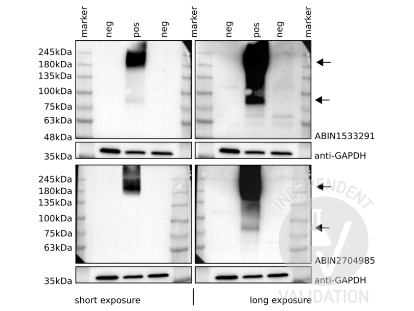
Passed. ABIN1533291 specifically recognizes human GRM6 expressed in CHO cells.
- Primary Antibody
- ABIN1533291
- Secondary Antibody
- HRP conjugated goat anti-rabbit antibody (Jackson Immuno Research, 111-035-144)
- Full Protocol
- Grow Chinese Hamster Ovary (CHO-K1) cells (ECACC) in DMEM (Sigma, D5671, lot RNB68272) supplemented with 10% Fetal calf Serum (Seraglob, S70500, lot 208/203142), 1x MEM Non-Essential-Amino-Acid (Sigma, M7145, lot RNBC8122), L-Alanyl-L-Glutammine (Merck, K0302) and Pen/Strep (Sigma, P4333, lot 058M4857V), at 37°C and 5% CO2.
- Plate 0.25x10⁶cells/ml in 2ml/well of cells in a 6 well plate.
- Grow cells for 24h at 37°C and 5% CO2.
- Transfect cells with 2ug/well of a plasmid expressing human GRM6 under the control of the ubiquitous promoter CMV using TransIt-LT1 transfection reagent (Mirus, Mir2300, lot 81104333) following the manufacturer´s instructions.
- Grow cells for 72h at 37°C and 5% CO2.
- Harvest cells with PBS and lyse them on ice for 30min with 50µl/well of RIPA buffer (25mM TrispH7-8, 150mM NaCl, 0.1% SDS, 0.5% sodium deoxycholate, 1% Triton X-100 or NP-40).
- Complete the lysis with a freeze/taw cycle.
- Determine total protein content of the lysates using Pierce BCA protein assay (Thermo Fisher, 23221).
- Denature 25μg of total protein for 20min at 37°C in 20μl Laemmli SDS sample buffer and subsequently separate them on a denaturing 4-20% Mini-PROTEAN TGX Stain-Free Gel (Bio-Rad, 456-8094) for 20min at 100V and 2h at 130V.
- Transfer proteins onto Immobilion-P transfer membrane (Immobilion, IPVH00010) for 75min at 100mA.
- Block the membrane with TBST 5% milk for 1h at RT.
- Incubate with primary
- rabbit anti-GRM6 antibody (antibodies-online, ABIN1533291, lot 310210) diluted 1:1000 in TBST 5% milk ON at 4°C,
- rabbit anti-GRM6 antibody (antibodies-online, ABIN2704985, lot 73482) diluted 1:1000 (positive control), or
- mouse anti-GAPDH antibody (Fitzgerald, 10R-G09a, lot 2417) diluted 1:40000 in TBST 5% milk ON at 4°C.
- Wash membrane 3x for 5min with TBST buffer.
- Incubation with secondary
- HRP conjugated goat anti-rabbit antibody (Jackson Immuno Research, 111-035-144) diluted 1:3000 in TBST 5% milk for 45min at RT or
- goat anti-mouse antibodies (Jackson Immuno Research, 115-035-146) diluted 1:3000 in TBST 5% milk for 45min at RT.
- Wash membrane 3x for 10min with TBST buffer.
- Reveal protein bands using Clarity MAX Western ECL Substrate (Bio-Rad, 1705062) on a ChemiDoc MP imaging system (Bio-Rad, 17001402).
- Experimental Notes
ABIN1533291 reveals two distinct bands. A lower one, which molecular weight corresponds to the monomer of the target protein, and a higher one which appears to correspond to a dimer of GRM6. The protein bands are only visible in the positive but not in the negative controls. Importantly, the same result was observed with another anti GRM6 antibody (ABIN2704985).
Being a G-Protein-Coupled Receptor it is not surprising that GRM6 forms strong dimers that can withstand boiling. Several attempts were performed in order to dissolve such dimers including sonication and different boiling protocols but they were unsuccessful. Similar results were obtained with HEK293 cells as well (not shown).
Validation #103490 (Western Blotting)
 Validation Images
![Western blot analysis of cell lysates from CHO-K1 cells transfected with a human GRM6 expression plasmid (pos) or untransfected cells (neg) using ABIN1533291 (top), ABIN2704985 (bottom) with short or long exposure times, or an anti-GAPDH loading control antibody. Arrows indicate what appear to be GRM6 monomers and dimers.]() Western blot analysis of cell lysates from CHO-K1 cells transfected with a human GRM6 expression plasmid (pos) or untransfected cells (neg) using ABIN1533291 (top), ABIN2704985 (bottom) with short or long exposure times, or an anti-GAPDH loading control antibody. Arrows indicate what appear to be GRM6 monomers and dimers.
Full Methods
-
- by
- Group Kleinlogel, Department of Physiology, University of Bern, Bern, Switzerland
- No.
- #103795
- Date
- 03/15/2019
- Antigen
- GRM6
- Lot Number
- 310210
- Method validated
- Immunofluorescence
- Positive Control
Chinese Hamster Ovary (CHO-K1) cells transfected with a plasmid expressing human GRM6 and turbo Red Fluorescent protein (tRFP)
- Negative Control
Non-transfected CHO-K1 cells
CHO transfected with a plasmid expressing human GRM6 stained with secondary antibody only
- Notes
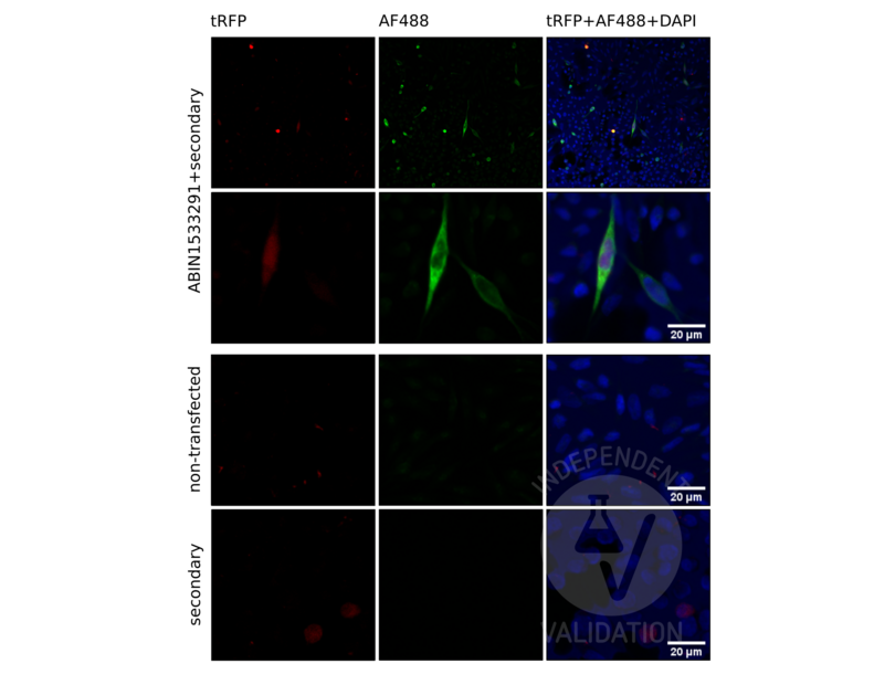
Passed. ABIN1533291 specifically labels the targeted antigen in transfected CHO-K1 cells in IF as demonstrated by the co-labeling with tRFP. No specific signal was detected in the negative controls.
- Primary Antibody
- ABIN1533291
- Secondary Antibody
- AF488 conjugated goat anti-rabbit IgG antibody (Invitrogen, A11008)
- Full Protocol
- Grow cells:
- Grow Chinese Hamster Ovary (CHO-K1) cells (ECACC) in DMEM (Sigma, D5671, lot RNB68272) supplemented with 10% Fetal calf Serum (Seraglob, S70500, lot 208/203142), 1x MEM Non-Essential-Amino-Acid (Sigma, M7145, lot RNBC8122), L-Alanyl-L-Glutammine (Merck, K0302) and Penicillin/Streptomycin (Sigma, P4333, lot 058M4857V) at 37°C and 5% CO2.
- Plate 0.125x10⁶cells/well in 0.5 ml/well in a 24 well plates on coverslips.
- Grow cells for 24h at 37°C and 5% CO2.
- Transfect cells with a plasmid expressing human GRM6-tRFP under the control of the CMV promoter using Polyethylenimine hydrochloride (PEI) reagent (Sigma, 764965):
- Add to 50μl of serum free Opti-MEM medium (Life Technologies, 31985-047) 0.5μg of DNA and 1.5μl of PEI.
- Add the mixture directly into the wells and incubate for 4-5h.
- Replace medium with fresh culture medium.
- Grow cells for 72h at 37°C and 5% CO2.
- Fixation:
- Fix cells in PFA 4% in PBS for 7-8min.
- Rinse 3x with 500µl 1x PBS.
- Add 200µl 0.1M Glycine in TBS for 15min.
- Remove Glycine with a pipette.
- Add blocking solution (5% Serum (NDS or NGS) + 1% BSA Serum in 0.3% Triton-X100 in 1x TBS) for 1h at RT.
- Primary antibody:
- Dilute primary rabbit anti-GRM6 antibody (antibodies-online, ABIN1533291, lot 310210) 1:100 in blocking solution.
- Spin the tube at 16000rcf for 6min to precipitate eventual particulate.
- Add 200µl primary antibody solution per well.
- Incubate slides in the dark ON at 4 °C.
- Wash slides 3x 10min with 500µl 0.3% Triton-X100 in 1x TBS.
- Secondary antibody:
- Dilute secondary AF488 conjugated goat anti-rabbit IgG antibody (Invitrogen, A11008) 1:400 and DAPI 1:2000 from a 1mg/ml stock dilution in blocking solution.
- Spin the tube at 16000rcf for 6min to precipitate eventual particulate.
- Add 200µl secondary antibody solution per well.
- Incubate slides in the dark for 1-2 h at RT.
- Wash slides 3x 10min with 500µl 0.3% Triton-X100 in 1x TBS.
- Mount slides:
- Mount slides with fluorescence mounting medium (DAKO, S3023, lot 10115314).
- Place a coverslip on it avoiding bubbles. Seal the edges of the coverslip with a clear nail polish.
- Let the nail polish dry and store samples at -4°C in the dark.
- Microscopy:
- Image acquisition on Zeiss LSM with a 20x objective. Gain(Master) of approximately 500.
- Experimental Notes
Validation #103795 (Immunofluorescence)
 Validation Images
![Low and high magnification pictures of CHO-K1 cells, transfected with a plasmid expressing human GRM6 and turbo Red Fluorescent protein (tRFP) stained with ABIN1533291 and an AF488 conjugated secondary antibody (AF488). Co-labeling of tRFP and GRM6 indicates the specificity of the antibody. Non-transfected cells (non-transfected) and transfected cells stained with the secondary antibody only (secondary) served as negative controls.]() Low and high magnification pictures of CHO-K1 cells, transfected with a plasmid expressing human GRM6 and turbo Red Fluorescent protein (tRFP) stained with ABIN1533291 and an AF488 conjugated secondary antibody (AF488). Co-labeling of tRFP and GRM6 indicates the specificity of the antibody. Non-transfected cells (non-transfected) and transfected cells stained with the secondary antibody only (secondary) served as negative controls.
Full Methods
-
-
Format
- Liquid
-
Concentration
- 1 mg/mL
-
Buffer
- Liquid in PBS containing 50 % glycerol, 0.5 % BSA and 0.02 % sodium azide.
-
Preservative
- Sodium azide
-
Precaution of Use
- This product contains Sodium azide: a POISONOUS AND HAZARDOUS SUBSTANCE which should be handled by trained staff only.
-
Storage
- -20 °C
-
Storage Comment
- Store at -20°C/1 year
-
Expiry Date
- 12 months
-
-
- Metabotropic Glutamate Receptor 6 (GRM6) (Glutamate Receptor, Metabotropic 6 (GRM6))
-
Alternative Name
- mGluR6
-
Background
-
Synonyms: GRM6, GPRC1F, MGLUR6, Metabotropic glutamate receptor 6, mGluR6
Background: glutamate metabotropic receptor 6(GRM6) Homo sapiens L-glutamate is the major excitatory neurotransmitter in the central nervous system and activates both ionotropic and metabotropic glutamate receptors. Glutamatergic neurotransmission is involved in most aspects of normal brain function and can be perturbed in many neuropathologic conditions. The metabotropic glutamate receptors are a family of G protein-coupled receptors, that have been divided into 3 groups on the basis of sequence homology, putative signal transduction mechanisms, and pharmacologic properties. Group I includes GRM1 and GRM5 and these receptors have been shown to activate phospholipase C. Group II includes GRM2 and GRM3 while Group III includes GRM4, GRM6, GRM7 and GRM8. Group II and III receptors are linked to the inhibition of the cyclic AMP cascade but differ in their agonist selectivities. [provided by RefSeq, Feb 2012],
Gene Name: GRM6
Protein Name: Metabotropic glutamate receptor 6
-
Molecular Weight
- 95 kDa
-
Gene ID
- 2916
-
OMIM
- 257270
-
UniProt
- O15303
Target
-