TGF beta Receptor 1 (AA 301-400) antibody
 (5 references)
 (1 validation)
Quick Overview for TGF beta Receptor 1 (AA 301-400) antibody (ABIN671256)
Target
Reactivity
Host
Clonality
Conjugate
Application
-
-
Binding Specificity
- AA 301-400
-
Cross-Reactivity
- Cow, Human, Mouse, Rat
-
Predicted Reactivity
- Cow
-
Purification
- Purified by Protein A.
-
Immunogen
- KLH conjugated synthetic peptide derived from human TGF-beta R1
-
Isotype
- IgG
-
-
anti-TGF beta Receptor 1 (AA 301-400) antibody (FITC)
Reactivity: Cow, Human, Mouse, Rat WB, IF (cc), IF (p) Host: Rabbit Polyclonal FITC
-
-
Application Notes
-
WB 1:300-5000
ELISA 1:500-1000
IHC-P 1:200-400
IHC-F 1:100-500
IF(IHC-P) 1:50-200
IF(IHC-F) 1:50-200
IF(ICC) 1:50-200 -
Restrictions
- For Research Use only
-
-
- by
- Molecular Pathology Core, University of Florida
- No.
- #029756
- Date
- 07/03/2014
- Antigen
- Lot Number
- 130902
- Method validated
- Immunohistochemistry
- Positive Control
- human normal colon, FFPE
- Negative Control
- human normal adipose, FFPE
- Notes
- Strong staining was observed in the positive control tissue sample. No staining was observed in the negative control tissue. Very light staining was observed in the isotype control and no staining was observed in the secondary-only control.
- Primary Antibody
- Antigen: TGF-beta Receptor I
- Catalog number: ABIN671256
- Lot number: 130902
- Secondary Antibody
- Antigen: Mach2 rabbit HRP-Polymer
- Lot number: 082813
- Full Protocol
- Human colon and human adipose (formalin fixed paraffin embedded) blocks were CTSI Tissue Bank in University of Florida.
- The 4 µm thick sections were treated by Citra in steamer for 20 min and cool down on the bench for another 20 min.
- Sections were blocked in sniper for 15 min at room temperature.
- Sections were incubated with primary antibody diluted 1:100 in antibody diluent for 1 h at room temperature.
- Sections were rinsed three times in TBS for 5 min each at RT.
- Sections were incubated with Mach2 rabbit HRP-Polymer for 30 min at RT.
- Sections were rinsed three times in TBS for 5 min each at RT.
- DAB color develop under light microscope.
- Coverslips were mounted on slides with Cytoseal XYL (Richard-Allan Scientific)
- Stained sections were imaged with a Zeiss Axioskop2 microscope.
- Experimental Notes
- - No experimental challenges noted.
Validation #029756 (Immunohistochemistry)
 Validation Images
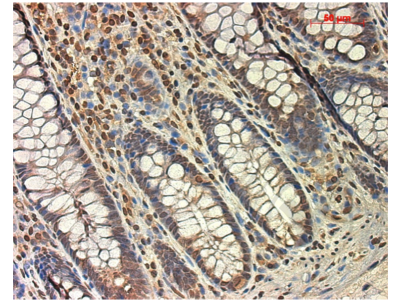
![Figure 1. Micrograph image of positive control (human colon tissue) stained with anti-TGF Beta R1 (brown staining). Scale bar = 50 µm.]() Figure 1. Micrograph image of positive control (human colon tissue) stained with anti-TGF Beta R1 (brown staining). Scale bar = 50 µm.
Figure 1. Micrograph image of positive control (human colon tissue) stained with anti-TGF Beta R1 (brown staining). Scale bar = 50 µm.
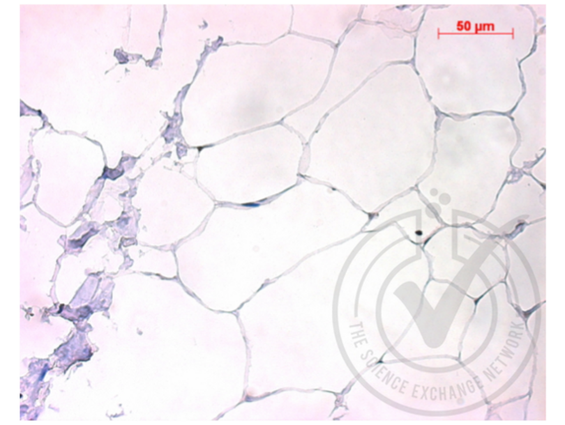
![Figure 2. Micrograph image of negative control (human adipose tissue) stained with anti-TGF Beta R1 (brown staining). Scale bar = 50 µm.]() Figure 2. Micrograph image of negative control (human adipose tissue) stained with anti-TGF Beta R1 (brown staining). Scale bar = 50 µm.
Figure 2. Micrograph image of negative control (human adipose tissue) stained with anti-TGF Beta R1 (brown staining). Scale bar = 50 µm.
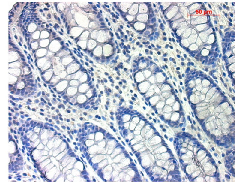
![Figure 3. Micrograph image of positive control (human colon tissue) stained with rabbit IgG isotype control antibody (brown staining). Scale bar = 50 µm.]() Figure 3. Micrograph image of positive control (human colon tissue) stained with rabbit IgG isotype control antibody (brown staining). Scale bar = 50 µm.
Figure 3. Micrograph image of positive control (human colon tissue) stained with rabbit IgG isotype control antibody (brown staining). Scale bar = 50 µm.
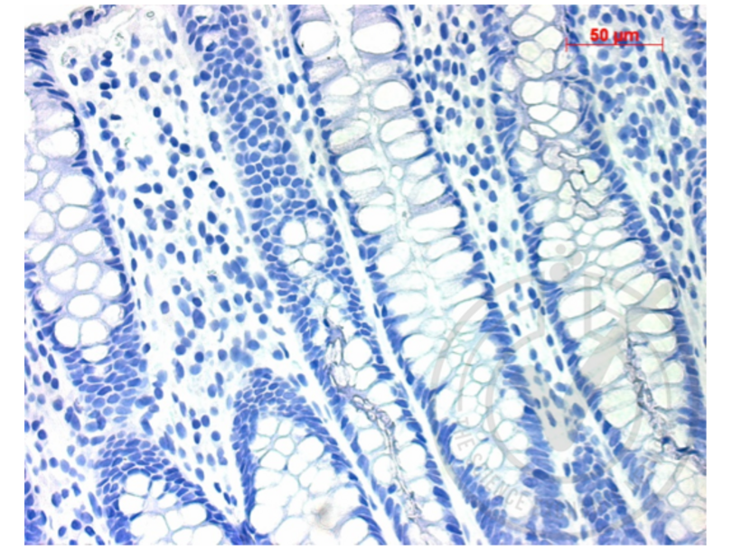
![Figure 4. Micrograph image of positive control (human colon tissue) stained with secondary antibody only (brown staining). Scale bar = 50 µm.]() Figure 4. Micrograph image of positive control (human colon tissue) stained with secondary antibody only (brown staining). Scale bar = 50 µm.
Full Methods
Figure 4. Micrograph image of positive control (human colon tissue) stained with secondary antibody only (brown staining). Scale bar = 50 µm.
Full Methods -
-
Format
- Liquid
-
Concentration
- 1 μg/μL
-
Buffer
- 0.01M TBS( pH 7.4) with 1 % BSA, 0.02 % Proclin300 and 50 % Glycerol.
-
Preservative
- ProClin
-
Precaution of Use
- This product contains ProClin: a POISONOUS AND HAZARDOUS SUBSTANCE, which should be handled by trained staff only.
-
Storage
- 4 °C,-20 °C
-
Storage Comment
- Shipped at 4°C. Store at -20°C for one year. Avoid repeated freeze/thaw cycles.
-
Expiry Date
- 12 months
-
-
-
: "GDF9 affects the development and tight junction functions of immature bovine Sertoli cells." in: Reproduction in domestic animals = Zuchthygiene, Vol. 52, Issue 4, pp. 640-648, (2017) (PubMed).
: "Regulation of cellular behaviors of fibroblasts related to wound healing by sol-gel derived bioactive glass particles." in: Journal of biomedical materials research. Part A, Vol. 104, Issue 10, pp. 2420-9, (2016) (PubMed).
: "Effect of iodine excess on Th1, Th2, Th17, and Treg cell subpopulations in the thyroid of NOD.H-2h4 mice." in: Biological trace element research, Vol. 159, Issue 1-3, pp. 288-96, (2014) (PubMed).
: "TNF-α enhances the effect of TGF-β on Gli2 expression in the KG-1 leukemic cell line." in: Experimental and therapeutic medicine, Vol. 8, Issue 2, pp. 676-680, (2014) (PubMed).
-
-
- TGF beta Receptor 1
-
Background
-
Synonyms: AAT5, ALK5, ESS1, LDS1, MSSE, SKR4, ALK-5, LDS1A, LDS2A, TGFR-1, ACVRLK4, TGF-beta receptor type-1, Activin A receptor type II-like protein kinase of 53kD, Activin receptor-like kinase 5, Serine/threonine-protein kinase receptor R4, TGF-beta type I receptor, Transforming growth factor-beta receptor type I, TGF-beta receptor type I, TbetaR-I, TGFBR1
Background: Transmembrane serine/threonine kinase forming with the TGF-beta type II serine/threonine kinase receptor, TGFBR2, the non-promiscuous receptor for the TGF-beta cytokines TGFB1, TGFB2 and TGFB3. Transduces the TGFB1, TGFB2 and TGFB3 signal from the cell surface to the cytoplasm and is thus regulating a plethora of physiological and pathological processes including cell cycle arrest in epithelial and hematopoietic cells, control of mesenchymal cell proliferation and differentiation, wound healing, extracellular matrix production, immunosuppression and carcinogenesis. The formation of the receptor complex composed of 2 TGFBR1 and 2 TGFBR2 Molecules symmetrically bound to the cytokine dimer results in the phosphorylation and the activation of TGFBR1 by the constitutively active TGFBR2. Activated TGFBR1 phosphorylates SMAD2 which dissociates from the receptor and interacts with SMAD4. The SMAD2-SMAD4 complex is subsequently translocated to the nucleus where it modulates the transcription of the TGF-beta-regulated genes. This constitutes the canonical SMAD-dependent TGF-beta signaling cascade. Also involved in non-canonical, SMAD-independent TGF-beta signaling pathways. For instance, TGFBR1 induces TRAF6 autoubiquitination which in turn results in MAP3K7 ubiquitination and activation to trigger apoptosis. Also regulates epithelial to mesenchymal transition through a SMAD-independent signaling pathway through PARD6A phosphorylation and activation.
-
Gene ID
- 7046
-
UniProt
- P36897
Target
-


