IDE antibody (AA 491-590)
 (2 validations)
Quick Overview for IDE antibody (AA 491-590) (ABIN723680)
Target
See all IDE AntibodiesReactivity
Host
Clonality
Conjugate
Application
-
-
Binding Specificity
- AA 491-590
-
Cross-Reactivity
- Human, Mouse, Rat
-
Predicted Reactivity
- Cow,Pig,Chicken
-
Purification
- Purified by Protein A.
-
Immunogen
- KLH conjugated synthetic peptide derived from human IDE
-
Isotype
- IgG
-
-
anti-Insulin-Degrading Enzyme (IDE) (AA 753-973) antibody
IDE Reactivity: Human WB, IHC Host: Rabbit Polyclonal unconjugated
anti-Insulin-Degrading Enzyme (IDE) (AA 1-250) antibodyIDE Reactivity: Human WB, IF Host: Rabbit Polyclonal unconjugated
anti-Insulin-Degrading Enzyme (IDE) (AA 920-1019) antibodyIDE Reactivity: Human WB, ELISA Host: Mouse Polyclonal unconjugated
anti-Insulin-Degrading Enzyme (IDE) (N-Term) antibodyIDE Reactivity: Human, Mouse, Rat WB, IHC, ELISA Host: Rabbit Polyclonal unconjugated
anti-Insulin-Degrading Enzyme (IDE) antibodyIDE Reactivity: Human WB, IF, IHC (p) Host: Mouse Monoclonal 1H4 unconjugated
anti-Insulin-Degrading Enzyme (IDE) (Internal Region) antibodyVerified IDE Reactivity: Human WB, IHC, ELISA Host: Goat Polyclonal unconjugated
anti-Insulin-Degrading Enzyme (IDE) antibodyKD Validated IDE Reactivity: Human WB, FACS Host: Rabbit Monoclonal 23GB6210 unconjugated Recombinant Antibody
anti-Insulin-Degrading Enzyme (IDE) (AA 501-800) antibodyIDE Reactivity: Human IHC, ELISA, IF Host: Rabbit Polyclonal unconjugated
anti-Insulin-Degrading Enzyme (IDE) antibodyIDE Reactivity: Human WB Host: Mouse Monoclonal 3H4 unconjugated
-
-
Application Notes
-
WB 1:300-5000
ELISA 1:500-1000
IHC-P 1:200-400
IHC-F 1:100-500
IF(IHC-P) 1:50-200
IF(IHC-F) 1:50-200
IF(ICC) 1:50-200 -
Restrictions
- For Research Use only
-
-
- by
- Prof. Merighi, Laboratory of Neurobiology, Department of Veterinary Sciences, University of Turin
- No.
- #104435
- Date
- 03/15/2023
- Antigen
- IDE
- Lot Number
- 9C07M588
- Method validated
- Immunohistochemistry
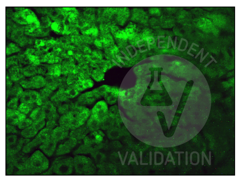
- Positive Control
Adult mouse liver fixed in 4% paraformaldehyde
- Negative Control
One control slice for each experimental procedure processed omitting the primary antibody; overnight incubation in diluent solution only.
- Notes
Passed. The IDE antibody (AA 491-590) ABIN723680 works in IHC-P at 1:100 concentrations with Tyramide amplification.
- Primary Antibody
- ABIN723680
- Secondary Antibody
- poly-HRP conjugated goat anti-rabbit antibody
- Full Protocol
- Perfuse mice with paraformaldehyde 4% in 0.1 M phosphate buffer pH 7.4 and post-fix in the same fixative for an additional 2 h at RT.
- Wash, dehydrate, and embed samples in paraffin wax.
- Wash several times with 0.01 M PBS.
- Cut liver with a microtome into 20 µm sections and mount on glass slides.
- After paraffin removal, incubate sections for 1 h at RT in PBS containing 1% albumin from chicken egg white (Sigma, A5378) and 0.3% Triton-X-100 (BioRad, 161-0407, lot 00583) to block non-specific binding sites.
- Incubate sections with primary rabbit anti-IDE (antibodies-online, ABIN723680, lot 9C07M588) diluted 1:50, 1:100, 1:200, and 1:300 in PBS-BSA-PLL ON at RT in a humid chamber.
- Wash sections 3x 5 min with 0.01 M PBS.
- Incubate sections with secondary poly-HRP conjugated goat anti-rabbit antibody from Alexa Fluor 488 Tyramide SuperBoost Kit, goat anti-rabbit IgG (Thermo Fisher Scientific, B40922, lot 2465062) for 1 h at RT.
- Wash sections 3x 5 min with 0.01 M PBS.
- Incubate sections with Tyramide working solution containing 100X Tyramide stock solution (Alexa 488), 100X H2O2 solution and 1X Reaction buffer for 10 min.
- Stop the reaction with the Reaction Stop Reagent working solution.
- Wash sections 3x 5 min with 0.01M PBS.
- Mount specimens in Fluoroshield (Sigma, F6182, lot MKCB0153V).
- Acquire images with a fluorescence microscope and appropriate filter settings for AF488, e.g. Leica DM 6000B fluorescence microscope equipped with a digital camera at 40x magnification.
- Experimental Notes
Validation #104435 (Immunohistochemistry)
 Validation Images
![Staining of IDE positive cells in the adult mouse liver with ABIN723680.]() Staining of IDE positive cells in the adult mouse liver with ABIN723680.
Full Methods
Staining of IDE positive cells in the adult mouse liver with ABIN723680.
Full Methods -
- by
- Prof. Merighi, Laboratory of Neurobiology, Department of Veterinary Sciences, University of Turin
- No.
- #104497
- Date
- 03/15/2023
- Antigen
- IDE
- Lot Number
- 9C07M588
- Method validated
- Western Blotting
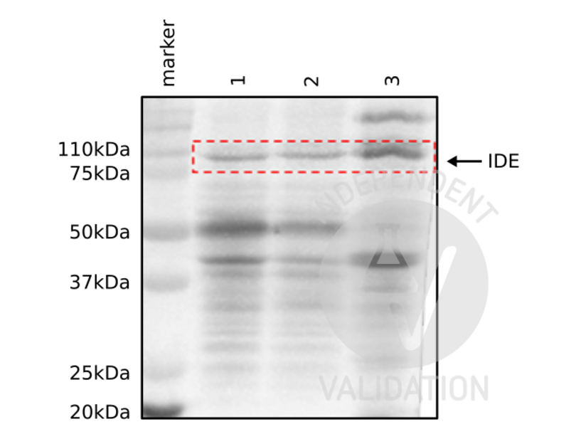
- Positive Control
Adult mouse brain, cerebellum, and liver
- Negative Control
- Notes
Passed. The IDE antibody (AA 491-590) ABIN723680 works in WB at 1:1000 concentrations with sensitive ECL substrate.
- Primary Antibody
- ABIN723680
- Secondary Antibody
- HRP-conjugated mouse anti-rabbit
- Full Protocol
- Homogenize tissues with cold lysis buffer containing 50 mM Tris HCl, 150 mM NaCl, 1% Triton X-100, 1 mM EDTA, and 1% protease inhibitor (Sigma P8340) using an ultrasonic homogenizer (MSE, SoniPrep 150) with 16 amplitude, 20 s on, 10 s off pulse for 60 s.
- Centrifuge tissue homogentates at 13,000 rpm for 20 min at 4 °C.
- Collect supernatants and Determine total protein content using a Bradford assay.
- Denature 50 µg of total protein for 5 min at 90 °C and subsequently separate them on a denaturing 12% PAGE-SDS gel alongside a Precision Plus Protein Dual Color Standard (Bio-Rad, 160374).
- Electro-transfer proteins onto nitrocellulose membrane (Amerscham Biosciences, RPN203D) ON in the cold room.
- Wash membrane 3x for 10 mon with 0.01 M PBS containing 0.1% Tween-20 (PBST).
- Block membrane with PBST containing 2% bovine serum albumin for 1 h at RT.
- Incubate membrane with primary rabbit anti-IDE antibody (antibodies-online, ABIN723680, lot 9C07M588) diluted 1:1,000 in PBST ON at 4 °C.
- Wash membrane 3x 10 min with PBST.
- Incubate membrane with secondary HRP-conjugated mouse anti-rabbit IgG (Sigma, A1949) diluted 1:4,000 in PBST for 1 h at RT.
- Wash membrane 3x 10 min with PBST.
- Visualize proteins with WesternBright Sirius HRP substrate (Advansta, K-12043) using a ChemiDoc Imaging System.
- Experimental Notes
Validation #104497 (Western Blotting)
 Validation Images
![Western blot detection of IDE (110 kDa) in adult mouse brain (1), cerebellum (2), and liver (3) tissue homogenates with ABIN723680.]() Western blot detection of IDE (110 kDa) in adult mouse brain (1), cerebellum (2), and liver (3) tissue homogenates with ABIN723680.
Full Methods
Western blot detection of IDE (110 kDa) in adult mouse brain (1), cerebellum (2), and liver (3) tissue homogenates with ABIN723680.
Full Methods -
-
Format
- Liquid
-
Concentration
- 1 μg/μL
-
Buffer
- 0.01M TBS( pH 7.4) with 1 % BSA, 0.02 % Proclin300 and 50 % Glycerol.
-
Preservative
- ProClin
-
Precaution of Use
- This product contains ProClin: a POISONOUS AND HAZARDOUS SUBSTANCE, which should be handled by trained staff only.
-
Storage
- 4 °C,-20 °C
-
Storage Comment
- Shipped at 4°C. Store at -20°C for one year. Avoid repeated freeze/thaw cycles.
-
Expiry Date
- 12 months
-
-
- IDE (Insulin-Degrading Enzyme (IDE))
-
Alternative Name
- IDE
-
Background
-
Synonyms: INSULYSIN, Insulin-degrading enzyme, Abeta-degrading protease, Insulin protease, Insulinase, IDE
Background: Plays a role in the cellular breakdown of insulin, IAPP, glucagon, bradykinin, kallidin and other peptides, and thereby plays a role in intercellular peptide signaling. Degrades amyloid formed by APP and IAPP. May play a role in the degradation and clearance of naturally secreted amyloid beta-protein by neurons and microglia.
-
Gene ID
- 3416
-
UniProt
- P14735
-
Pathways
- SARS-CoV-2 Protein Interactome
Target
-


