Ki-67 ELISA Kit
 (2 references)
 (1 validation)
Quick Overview for Ki-67 ELISA Kit (ABIN415150)
Target
See all Ki-67 (MKI67) ELISA KitsReactivity
Detection Method
Method Type
Detection Range
Application
Sample Type
-
-
Minimum Detection Limit
- 0.78 ng/mL
-
Purpose
-
The kit is a sandwich enzyme immunoassay for in vitro quantitative measurement of Ki67P in human tissue homogenates, cell lysates, cell culture supernates.
We offer validation data (WB) for the kit components. So you can be sure to order a reliable ELISA kit product composed of high quality reagents. -
Analytical Method
- Quantitative
-
Specificity
-
This assay has high sensitivity and excellent specificity for detection of Ki-67 Protein (Ki-67).
No significant cross-reactivity or interference between Ki-67 Protein (Ki-67) and analogues was observed. -
Cross-Reactivity (Details)
- No significant cross-reactivity or interference between Ki-67 Protein (Ki67P) and analogues was observed.
-
Sensitivity
- < 0.32 ng/mL
-
Components
-
- Pre-coated, 96-well strip plate
- Plate sealer
- Standard
- Standard Diluent
- Detection Reagent A
- Assay Diluent A
- Detection Reagent B
- Assay Diluent B
- TMB Substrate
- Stop Solution
- Wash Buffer (30X)
- Instruction manual
-
Material not included
-
- Microplate reader with 450 nm filter.
- Precision single or multi-channel pipettes and disposable tips.
- Eppendorf Tubes for diluting samples.
- Deionized or distilled water.
- Absorbent paper for blotting the microtiter plate.
- Container for Wash Solution
-
-
Antigen Identified By Monoclonal Antibody Ki-67 (MKI67) ELISA Kit
MKI67 Reactivity: Human Colorimetric Sandwich ELISA 0.78 ng/mL - 50 ng/mL Cell Culture Supernatant, Cell Lysate, Tissue Homogenate
Antigen Identified By Monoclonal Antibody Ki-67 (MKI67) ELISA KitMKI67 Reactivity: Rat Colorimetric Sandwich ELISA 0.156 ng/mL - 10 ng/mL Cell Lysate, Plasma, Serum, Tissue Homogenate
Antigen Identified By Monoclonal Antibody Ki-67 (MKI67) ELISA KitMKI67 Reactivity: Mouse Colorimetric Sandwich ELISA 0.313 ng/mL - 20 ng/mL Plasma, Serum, Tissue Homogenate
Antigen Identified By Monoclonal Antibody Ki-67 (MKI67) ELISA KitMKI67 Reactivity: Human Colorimetric Sandwich ELISA 0.313 ng/mL - 20 ng/mL Plasma, Serum, Tissue Homogenate
Antigen Identified By Monoclonal Antibody Ki-67 (MKI67) ELISA KitMKI67 Reactivity: Mouse Colorimetric Sandwich ELISA 0.31 ng/mL - 20 ng/mL Cell Culture Supernatant, Cell Lysate, Tissue Homogenate
Antigen Identified By Monoclonal Antibody Ki-67 (MKI67) ELISA KitMKI67 Reactivity: Rat Colorimetric Sandwich ELISA 0.31 ng/mL - 20 ng/mL Cell Culture Supernatant, Cell Lysate, Tissue Homogenate
Antigen Identified By Monoclonal Antibody Ki-67 (MKI67) ELISA KitWide Range MKI67 Reactivity: Human Colorimetric Sandwich ELISA 0.78 ng/mL - 50 ng/mL Cell Culture Supernatant, Cell Lysate, Tissue Homogenate
Antigen Identified By Monoclonal Antibody Ki-67 (MKI67) ELISA KitMKI67 Reactivity: Mouse Colorimetric Sandwich ELISA 0.31 ng/mL - 20 ng/mL
Antigen Identified By Monoclonal Antibody Ki-67 (MKI67) ELISA KitMKI67 Reactivity: Human Colorimetric Sandwich ELISA 0.313 ng/mL - 20 ng/mL Plasma, Serum, Tissue Homogenate
Antigen Identified By Monoclonal Antibody Ki-67 (MKI67) ELISA KitMKI67 Reactivity: Human Colorimetric Sandwich ELISA 312 pg/mL - 20000 pg/mL Cell Culture Supernatant, Cell Lysate, Plasma, Serum, Tissue Homogenate
-
-
Application Notes
-
- Limited by the current condition and scientific technology, we cannot completely conduct the comprehensive identification and analysis on the raw material provided by suppliers. So there might be some qualitative and technical risks to use the kit.
- The final experimental results will be closely related to validity of the products, operation skills of the end users and the experimental environments. Please make sure that sufficient samples are available.
- Kits from different batches may be a little different in detection range, sensitivity and color developing time.
- Do not mix or substitute reagents from one kit lot to another. Use only the reagents supplied by manufacturer.
- Protect all reagents from strong light during storage and incubation. All the bottle caps of reagents should be covered tightly to prevent the evaporation and contamination of microorganism.
- There may be some foggy substance in the wells when the plate is opened at the first time. It will not have any effect on the final assay results. Do not remove microtiter plate from the storage bag until needed.
- Wrong operations during the reagents preparation and loading, as well as incorrect parameter setting for the plate reader may lead to incorrect results. A microplate plate reader with a bandwidth of 10nm or less and an optical density range of 0-3 O.D. or greater at 450 ± 10nm wavelength is acceptable for use in absorbance measurement. Please read the instruction carefully and adjust the instrument prior to the experiment.
- Even the same operator might get different results in two separate experiments. In order to get better reproducible results, the operation of every step in the assay should be controlled. Furthermore, a preliminary experiment before assay for each batch is recommended.
- Each kit has been strictly passed Q.C test. However, results from end users might be inconsistent with our in-house data due to some unexpected transportation conditions or different lab equipments. Intra-assay variance among kits from different batches might arise from above factors, too.
- Kits from different manufacturers for the same item might produce different results, since we have not compared our products with other manufacturers.
-
Comment
-
Information on standard material:
The standard might be recombinant protein or natural protein, that will depend on the specific kit. Moreover, the expression system is E.coli or yeast or mammal cell. There is 0.05% proclin 300 in the standard as preservative.
Information on reagents:
The stop solution used in the kit is sulfuric acid with concentration of 1 mol/L. And the wash solution is TBS. The standard diluent contains 0.02 % sodium azide, assay diluent A and assay diluent B contain 0.01% sodium azide. Some kits can contain is BSA in them.
Information on antibodies:
The provided antibodies and their host vary in different kits. -
Sample Volume
- 100 μL
-
Assay Time
- 3 h
-
Plate
- Pre-coated
-
Protocol
-
1. Prepare all reagents, samples and standards;
2. Add 100µL standard or sample to each well. Incubate 2 hours at 37°C;
3. Aspirate and add 100µL prepared Detection Reagent A. Incubate 1 hour at 37°C;
4. Aspirate and wash 3 times;
5. Add 100µL prepared Detection Reagent B. Incubate 30 minutes at 37°C;
6. Aspirate and wash 5 times;
7. Add 90µL Substrate Solution. Incubate 10-20 minutes at 37°C;
8. Add 50µL Stop Solution. Read at 450nm immediately.
-
Assay Precision
-
Intra-assay Precision (Precision within an assay): 3 samples with low, middle and high level Ki-67 Protein (Ki-67) were tested 20 times on one plate, respectively.
Inter-assay Precision (Precision between assays): 3 samples with low, middle and high level Ki-67 Protein (Ki-67) were tested on 3 different plates, 8 replicates in each plate.
CV(%) = SD/meanX100
Intra-Assay: CV<10%
Inter-Assay: CV<12%
-
Restrictions
- For Research Use only
-
-
- by
- Alamo Laboratories
- No.
- #029832
- Date
- 02/28/2015
- Antigen
- Lot Number
- L141016326
- Method validated
- ELISA
- Positive Control
- MCF7 lysate
- Negative Control
- Mouse brain lysate
- Notes
- Target protein was detected in the positive control sample and not in the negative control sample as expected.
- Primary Antibody
- Secondary Antibody
- Full Protocol
- 1. All reagents in the ELISA kit were brought up to room temperature (RT) before use.
- 2. 100 μL of standard or sample were added to wells in ELISA plate pre-coated with capture antibody.
- 3. All samples and standards were assayed in triplicate.
- 4. The plate was covered with sealer (provided in kit) and incubated for 2 hours at 37°C. Unbound material was aspirated but the wells were NOT washed.
- 5. 100 μL of Detection Reagent-A Working Solution was added to each well. Plate was covered with sealer (provided in kit) and incubated for 1 hour at 37°C. Unbound material was removed from each well and plate was washed three times with 350 μL of 1x Wash Solution (provided in the kit). After the last wash the plate was inverted and blotted against clean absorbent paper to remove any remaining liquid.
- 6. 100 μL of Detection Reagent-B Working Solution was added to each well. Plate was covered with sealer (provided in kit) and incubated for 30 minutes at 37°C.
- 7. Unbound material was removed by washing five times with 350 μL of 1x Wash Solution (provided in the kit). After the last wash the plate was inverted and blotted against clean absorbent paper to remove any remaining liquid.
- 8. 90 μL of Substrate Solution was added to wells and the plate was covered with a new plate sealer. The plate was gently tapped to ensure mixing and incubated for 25 minutes at 37°C in the dark.
- 9. After 25 minutes, when an apparent gradient appeared in the standard wells, the reaction was terminated by adding 50 μL of Stop Solution to each well.
- 10. The optical density (OD value) of each well was read using a microplate reader set to 450 nm.
- 11. The triplicate readings for each sample were averaged and the average zero standard optical density subtracted to yield ‘corrected absorbance at 450 nm’. A standard curve was generated by plotting the mean OD value for each standard on the X-axis against the concentration on the Y-axis using Excel. Standard curve was generated by regression analysis with three-parameter logistic.
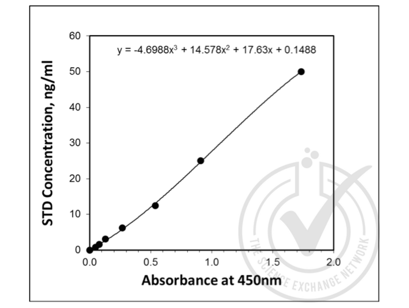
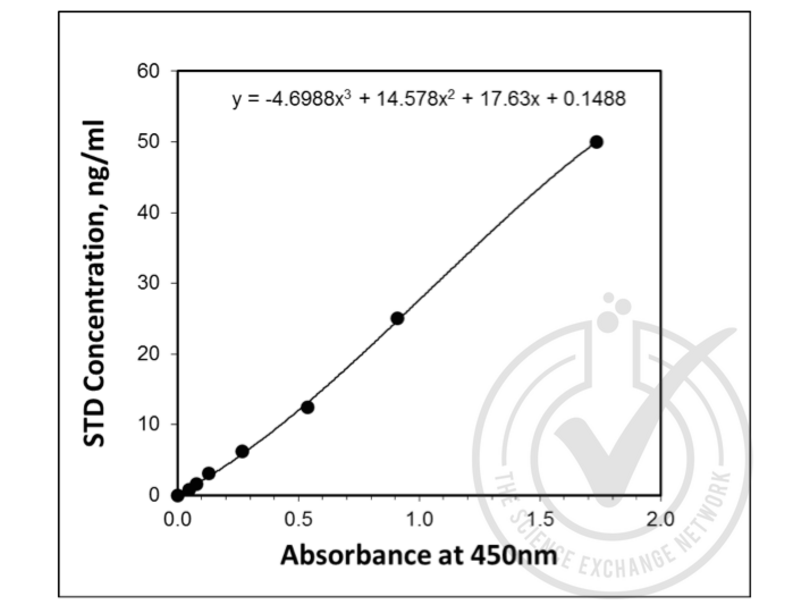
- 12. An equation (y = -4.6988x^3 + 14.578x^2 + 17.63x + 0.1488) was derived from the standard curve and used to calculate Ki67P concentrations in samples based on their Average Absorbance values.
- Experimental Notes
- No experimental challenges noted.
Validation #029832 (ELISA)
 Validation Images
![Figure 1: Graph of ‘Corrected’ OD450 nm plotted for standard curve samples. Standard curve was generated by regression analysis with three-parameter logistic. An equation (y = -4.6988x^3 + 14.578x^2 + 17.63x + 0.1488) was derived from the standard curve and used to calculate KI67P concentrations shown in Figure 2.]() Figure 1: Graph of ‘Corrected’ OD450 nm plotted for standard curve samples. Standard curve was generated by regression analysis with three-parameter logistic. An equation (y = -4.6988x^3 + 14.578x^2 + 17.63x + 0.1488) was derived from the standard curve and used to calculate KI67P concentrations shown in Figure 2.
Figure 1: Graph of ‘Corrected’ OD450 nm plotted for standard curve samples. Standard curve was generated by regression analysis with three-parameter logistic. An equation (y = -4.6988x^3 + 14.578x^2 + 17.63x + 0.1488) was derived from the standard curve and used to calculate KI67P concentrations shown in Figure 2.
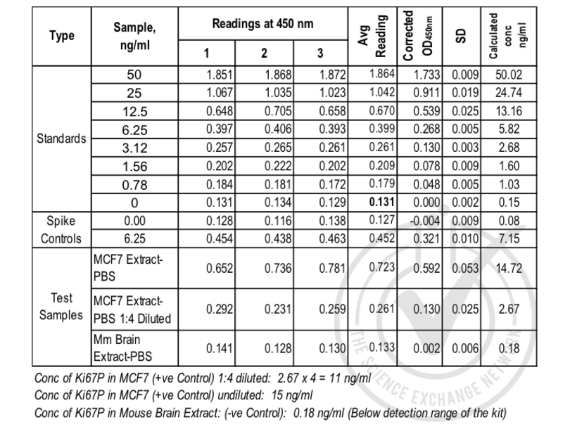
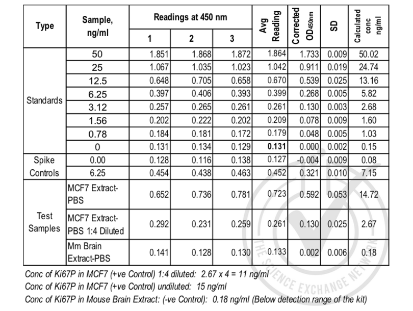
![Figure 2: Table of absorbance values for standard curve, spike control and samples.
Value for Avg Reading is derived from the average reading of three samples. Avg Reading for “0” amount of Standard was subtracted from all Avg Readings to yield “Corrected OD450 nm values” for Standards, spike controls and unknown samples. Standard deviation is included for all samples.]() Figure 2: Table of absorbance values for standard curve, spike control and samples.
Value for Avg Reading is derived from the average reading of three samples. Avg Reading for “0” amount of Standard was subtracted from all Avg Readings to yield “Corrected OD450 nm values” for Standards, spike controls and unknown samples. Standard deviation is included for all samples.
Full Methods
Figure 2: Table of absorbance values for standard curve, spike control and samples.
Value for Avg Reading is derived from the average reading of three samples. Avg Reading for “0” amount of Standard was subtracted from all Avg Readings to yield “Corrected OD450 nm values” for Standards, spike controls and unknown samples. Standard deviation is included for all samples.
Full Methods -
-
Precaution of Use
- The Stop Solution suggested for use with this kit is an acid solution. Wear eye, hand, face, and clothing protection when using this material.
-
Handling Advice
-
The stability of kit is determined by the loss rate of activity. The loss rate of this kit is less than 5 % within the expiration date under appropriate storage condition.
To minimize extra influence on the performance, operation procedures and lab conditions, especially room temperature, air humidity, incubator temperature should be strictly controlled. It is also strongly suggested that the whole assay is performed by the same operator from the beginning to the end. -
Storage
- 4 °C
-
Storage Comment
-
- For unopened kit: All the reagents should be kept according to the labels on vials. The Standard, Detection Reagent A, Detection Reagent B and the 96-well strip plate should be stored at -20°C upon receipt while the others should be at 4°C.
- For opened kit: When the kit is opened, the remaining reagents still need to be stored according to the above storage condition. Besides, please return the unused wells to the foil pouch containing the desiccant pack, and reseal along entire edge of zip-seal.
Note: It is highly recommended to use the remaining reagents within 1 month provided this is within the expiration date of the kit. - For ELISA kit, 1 day storage at 37°C can be considered as 2 months at 4°C, which means 3 days at 37°C equaling 6 months at 4°C.
-
Expiry Date
- 6 months
-
-
-
: "Cytotoxic and Apoptotic Effects of Luffa Cylindrica Leaves Extract against Acute Lymphoblastic Leukemic Stem Cells." in: Asian Pacific journal of cancer prevention : APJCP, Vol. 21, Issue 12, pp. 3661-3668, (2021) (PubMed).
: "Chemical Composition of Aqueous Ethanol Extract of Luffa cylindrica Leaves and Its Effect on Representation of Caspase-8, Caspase-3, and the Proliferation Marker Ki67 in Intrinsic Molecular Subtypes ..." in: Chemistry & biodiversity, Vol. 15, Issue 8, pp. e1800045, (2018) (PubMed).
-
-
- Ki-67 (MKI67) (Antigen Identified By Monoclonal Antibody Ki-67 (MKI67))
-
Alternative Name
- Ki67P
-
Background
- MKI67, KIA, Ki67P, Antigen Identified By Monoclonal Antibody Ki-67
-
UniProt
- P46013
-
Pathways
- Glycosaminoglycan Metabolic Process
Target See all Ki-67 (MKI67) ELISA Kits
-