p53 antibody (meLys370)
 (1 validation)
Quick Overview for p53 antibody (meLys370) (ABIN4902067)
Target
See all p53 (TP53) AntibodiesReactivity
Host
Clonality
Conjugate
Application
-
-
Binding Specificity
- meLys370
-
Purpose
- Rabbit polyclonal to p53 (Mono Methyl Lys370).
-
Specificity
- Mono-Methyl-p53 (K370) Polyclonal Antibody detects endogenous levels of p53 around the methylation site of K370 protein.
-
Characteristics
- Rabbit Polyclonal to p53 (Mono Methyl Lys370).
-
Purification
- The antibody was affinity-purified from rabbit antiserum by affinity-chromatography using epitope-specific immunogen.
-
Immunogen
- Synthesized peptide derived from human p53 around the methylation site of K370.
-
Isotype
- IgG
-
-
anti-Tumor Protein P53 (TP53) (pSer392) antibody
TP53 Reactivity: Human, Mouse, Rat WB, IHC, ELISA, IF, ICC Host: Rabbit Polyclonal unconjugated
anti-Tumor Protein P53 (TP53) (N-Term) antibodyTP53 Reactivity: Human WB, IF, IHC (p), IP, ICC Host: Rabbit Polyclonal unconjugated
anti-Tumor Protein P53 (TP53) antibodyKO Validated TP53 Reactivity: Human WB, IF, IHC (p), IP, ICC, ChIP Host: Rabbit Polyclonal unconjugated
anti-Tumor Protein P53 (TP53) antibodyTP53 Reactivity: Human, Mouse, Rat, Monkey WB, ELISA, IF, IHC (p) Host: Rabbit Polyclonal unconjugated
anti-Tumor Protein P53 (TP53) (AA 301-393) antibodyTP53 Reactivity: Human, Mouse, Rat, Goat WB, ELISA, FACS, IHC (p), ICC, IF (p), IF (cc), IHC (fro) Host: Rabbit Polyclonal unconjugated
anti-Tumor Protein P53 (TP53) (pSer20) antibodyTP53 Reactivity: Human, Mouse, Rat WB, IHC, ELISA, IF, ICC Host: Rabbit Polyclonal unconjugated
anti-Tumor Protein P53 (TP53) (acLys319) antibodyTP53 Reactivity: Human, Mouse, Rat WB, IHC, ELISA, IF, ICC Host: Rabbit Polyclonal unconjugated
anti-Tumor Protein P53 (TP53) (pSer15) antibodyTP53 Reactivity: Human, Mouse, Rat WB, IHC, ELISA, IF, IP, ICC Host: Rabbit Polyclonal unconjugated
anti-Tumor Protein P53 (TP53) (AA 293-322) antibodyTP53 Reactivity: Human WB, IF, IHC (p) Host: Rabbit Polyclonal RB7777 unconjugated
anti-Tumor Protein P53 (TP53) (AA 94-201) antibodyTP53 Reactivity: Human WB, ELISA, IF, IHC (p), IP Host: Mouse Monoclonal 2C3 unconjugated
-
-
Application Notes
-
WB 1:500-1:2000
ELISA 1:10000 -
Comment
-
Ubiquitous. Isoforms are expressed in a wide range of normal tissues but in a tissue-dependent manner. Isoform 2 is expressed in most normal tissues but is not detected in brain, lung, prostate, muscle, fetal brain, spinal cord and fetal liver. Isoform 3 is expressed in most normal tissues but is not detected in lung, spleen, testis, fetal brain, spinal cord and fetal liver. Isoform 7 is expressed in most normal tissues but is not detected in prostate, uterus, skeletal muscle and breast. Isoform 8 is detected only in colon, bone marrow, testis, fetal brain and intestine. Isoform 9 is expressed in most normal tissues but is not detected in brain, heart, lung, fetal liver, salivary gland, breast or intestine.
-
Restrictions
- For Research Use only
-
-
- by
- Department of Prevention and Therapy of Chronic Diseases, Institute for Advanced Biosciences, CNRS UMR5309
- No.
- #101268
- Date
- 08/03/2017
- Antigen
- p53K370me
- Lot Number
- B1001
- Method validated
- Western Blotting
- Positive Control
p53, in vitro specifically p53K370me1 methylated using methyltransferase SMYD2
ectopic SMYD-methylated wildtype p53 from 293T cell extracts
- Negative Control
p53, in vitro methylated using methyltransferases G9a and SETD8
p53ΔCTD, lacking C-terminal domain containing known methylable lysines
p53K370R, lacking the lysine 370 methylation site, mutated into arginine
- Notes
Passed. ABIN4902067 specifically recognizes monomethylated p53K370me1 and to a lesser extent p53Kme1.
- Primary Antibody
- ABIN4902067
- Secondary Antibody
- HRP-conjugated goat anti-rabbit or anti-mouse antibody (Biorad)
- Full Protocol
- In vitro p53 methylation assay:
- Clone SMYD2 and p53 from human cDNA library into pGex6p1 vector.
- Perform p53 mutagenesis using QuickChange II site-directed mutagenesis kit (Agilent, 200523).
- Transform vectors into BL21 pLys bacteria (Agilent) and induced protein expression with 0.1mM IPTG in 250mL culture ON at 19°C.
- Purify proteins by GST pulldown and quantify using Bradford assay and Coomassie gel staining.
- For the in vitro methylation assays incubate 1 to 2µg recombinant protein with 1µg of recombinant methyltransferases and 2µCi 3H-AdoMet (American Radiolabelled Chemicals) in buffer containing 50mM Tris-HCl pH8.0, 10% glycerol, 20mM KCl, 5mM MgCl2, and 1mMPMSF at ON at 30°C.
- Resolve the reaction mixture by SDS–PAGE, followed by autoradiography and Coomassie stain (Pierce).
- "In cellulo” p53 methylation assay:
- Subclone SMYD2 and p53 constructs from pGex6p1 vectors into pcagFLAG (SMYD2) or pCDNA3.1HA (p53).
- Transfect plasmids into 293T cells using Lipofectamine 2000 (ThermoFisher) according to supplier’s protocol.
- Harvest cells 36h post-transfection to allow methylation of ectopic p53 by SMYD2 in cells.
- Western blot:
- Separate 20µg of each sample on freshly cast SDS-PAGE gel in a gel electrophoresis chamber (BioRad) for 1h at 140V and RT.
- Transfer proteins onto ethanol-activated 0.2µm Hybond PVDF membrane (Amersham) with a semi-dry Trans-Blot Turbo (BioRad) for 30min at 20V.
- Block the membrane using 5% milk in TBS-Tween 0.1% for 1h at RT.
- Incubate the membrane with primary rabbit anti-p53 meLys370 antibody (antibodies-online, ABIN4902067, LOT B1001), mouse anti-HA antibody (Sigma), or mouse anti-Flag tag antibody (Sigma) diluted 1:1000 in 1% BSA TBS-Tween 0.1% ON at 4°C.
- Wash the membrane 3x for 10min in TBS-Tween 0.1%.
- Incubate the membrane with secondary HRP-conjugated goat anti-rabbit or anti-mouse antibody (Biorad) diluted 1:3000 in 1% BSA TBS-Tween 0.1% for 1h at RT.
- Wash the membrane 3x for 10min in TBS-Tween 0.1%.
- Visualize protein bands using ECL RevelBlot intense (Ozyme) and signal detection on Hyperfilm ECL 18 x24cm (Amersham).
- Experimental Notes
- Antibody ABIN4902067 recognizes greatly its specific substrate in vitro: it shows a strong signal when SMYD2 is present, implying monomethylation of K370. The signal is lost when using p53K370R and p53ΔCTD mutants. However, ABIN4902067 also recognizes to a lesser extent in vitro other p53 methylation such as p53 K373me1/2 and p53K382me2 caused by G9a and SETD8 respectively.
- The antibody does recognize unmethylated p53 when using a large quantity of the recombinant protein (not shown).
- ABIN4902067 gives a very clean and specific signal when using ectopic p53 from 293T cell extract: only p53 methylated by SMYD2 is detected, not the p53K370R mutant. The antibody can even be used without an initial immunoprecipitation of total p53 as observable in our figure.
- The binding affinity of ABIN4902067 can be ordered as follows: p53K370me1>p53Kme1>>>p53.
Validation #101268 (Western Blotting)
 Validation Images
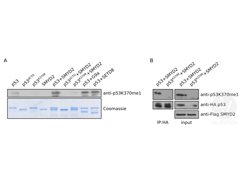
![A. In vitro methylation assay of wt p53 and the p53K370R and p53ΔCTD mutants by methyltransferases SMYD2, G9a, and SETD8 (upper panel). See experimental notes for details. The lower panel shows a Commassie stain of the gel prior to the immunoblot. B. Western blot of 293T cellular extracts ectopically expressing HA-tag wt p53 or p53K370R in the presence of DYKDDDDK-tagged SMYD2, verified using an DYKDDDDK-tag antibody. In the panels on the left p53 in the lysates was enriched using an HA-tag antibody. WB with an HA-tag antibody reveals immunoprecipitated wt p53 and p53K370R whereas ABIN4902067 reveals specifically SMYD2-methylated wt p53.]() A. In vitro methylation assay of wt p53 and the p53K370R and p53ΔCTD mutants by methyltransferases SMYD2, G9a, and SETD8 (upper panel). See experimental notes for details. The lower panel shows a Commassie stain of the gel prior to the immunoblot. B. Western blot of 293T cellular extracts ectopically expressing HA-tag wt p53 or p53K370R in the presence of DYKDDDDK-tagged SMYD2, verified using an DYKDDDDK-tag antibody. In the panels on the left p53 in the lysates was enriched using an HA-tag antibody. WB with an HA-tag antibody reveals immunoprecipitated wt p53 and p53K370R whereas ABIN4902067 reveals specifically SMYD2-methylated wt p53.
Full Methods
A. In vitro methylation assay of wt p53 and the p53K370R and p53ΔCTD mutants by methyltransferases SMYD2, G9a, and SETD8 (upper panel). See experimental notes for details. The lower panel shows a Commassie stain of the gel prior to the immunoblot. B. Western blot of 293T cellular extracts ectopically expressing HA-tag wt p53 or p53K370R in the presence of DYKDDDDK-tagged SMYD2, verified using an DYKDDDDK-tag antibody. In the panels on the left p53 in the lysates was enriched using an HA-tag antibody. WB with an HA-tag antibody reveals immunoprecipitated wt p53 and p53K370R whereas ABIN4902067 reveals specifically SMYD2-methylated wt p53.
Full Methods -
-
Format
- Liquid
-
Concentration
- 1 mg/mL
-
Buffer
- Liquid in PBS containing 50 % glycerol, 0.5 % BSA and 0.02 % sodium azide.
-
Preservative
- Sodium azide
-
Precaution of Use
- This product contains Sodium azide: a POISONOUS AND HAZARDOUS SUBSTANCE which should be handled by trained staff only.
-
Handling Advice
- Avoid repeated freeze/thaw cycles.
-
Storage
- -20 °C
-
Storage Comment
- Store at -20°C, and avoid repeat freeze-thaw cycles.
-
-
- p53 (TP53) (Tumor Protein P53 (TP53))
-
Alternative Name
- p53
-
Gene ID
- 7157
-
UniProt
- P04637
-
Pathways
- p53 Signaling, MAPK Signaling, PI3K-Akt Signaling, Apoptosis, AMPK Signaling, Chromatin Binding, ER-Nucleus Signaling, Positive Regulation of Endopeptidase Activity, Hepatitis C, Protein targeting to Nucleus, Autophagy, Warburg Effect
Target
-


