SYNGAP1 antibody (Internal Region)
 (1 validation)
-
- Target See all SYNGAP1 Antibodies
- SYNGAP1 (Synaptic Ras GTPase Activating Protein 1 (SYNGAP1))
-
Binding Specificity
- AA 1169-1183, Internal Region
-
Reactivity
- Human, Mouse, Rat, Cow, Dog, Pig
-
Host
- Goat
-
Clonality
- Polyclonal
-
Conjugate
- This SYNGAP1 antibody is un-conjugated
-
Application
- Western Blotting (WB), Enzyme Immunoassay (EIA)
- Specificity
- This antibody reacts to SYNGAP1.
- Cross-Reactivity (Details)
-
Species reactivity (expected):Human, Rat, Canine, Porcine and Bovine.
Species reactivity (tested):Mouse. - Purification
- Affinity Purified
- Immunogen
- Peptide with sequence C-ESAHIEREEYKLKEY, from the internal region (near C Terminus) of the protein sequence according to NP_006763.2.
- Top Product
- Discover our top product SYNGAP1 Primary Antibody
-
anti-Synaptic Ras GTPase Activating Protein 1 (SYNGAP1) (AA 1161-1343) antibody
SYNGAP1 Reactivity: Human ELISA, IF Host: Rabbit Polyclonal unconjugated
anti-Synaptic Ras GTPase Activating Protein 1 (SYNGAP1) (C-Term) antibodySYNGAP1 Reactivity: Human, Mouse, Rat WB, ELISA Host: Rabbit Polyclonal unconjugated
anti-Synaptic Ras GTPase Activating Protein 1 (SYNGAP1) (AA 1169-1183) antibodyVerified SYNGAP1 Reactivity: Human, Mouse WB, ELISA, IF, IHC Host: Goat Polyclonal unconjugated
anti-Synaptic Ras GTPase Activating Protein 1 (SYNGAP1) (AA 60-72) antibodyVerified SYNGAP1 Reactivity: Mouse WB, ELISA Host: Goat Polyclonal unconjugated
anti-Synaptic Ras GTPase Activating Protein 1 (SYNGAP1) antibodySYNGAP1 Reactivity: Human WB Host: Rabbit Monoclonal unconjugated
anti-Synaptic Ras GTPase Activating Protein 1 (SYNGAP1) (AA 1185-1199), (C-Term) antibodySYNGAP1 Reactivity: Human, Mouse, Rat WB, IF, IHC Host: Rabbit Polyclonal unconjugated
-
- Application Notes
- Optimal working dilution should be determined by the investigator.
- Restrictions
- For Research Use only
-
- by
- Molekulare Neurobiologie, Max-Planck-Institut für Experimentelle Medizin
- No.
- #100923
- Date
- 04/08/2017
- Antigen
- SYNGAP1
- Lot Number
- P1
- Method validated
- Western Blotting
- Positive Control
- SYNGAP1 transfected HEK293TF cells, anti-SYNGAP1 antibody PA1-046
- Negative Control
- untransfected HEK293TF cells
- Notes
- Passed. ABIN955030 specifically detects SynGAP in transiently transfected HEK293TF cells expressing murine SynGAP.
- Primary Antibody
- ABIN955030
- Secondary Antibody
- mouse anti-goat HRP-conjugate (Sigma)
- Full Protocol
- HEK293TF cells are grown in in 10 cm cell culture dishes in DMEM (1x) + Glutamax TM-I (Gibco, 31966-021, lot 1838117) supplemented with 10% fetal bovine serum Gibco, 105000064, lot 08F7660K) and 0.5% Penicillin/Streptomycin (Gibco, 15140122, lot 1864845)at 37°C in 5% CO2.
- Transfect cells at 50% confluence with plasmid encoding full-length murine SynGAP or empty pcDNA3(+) vector as negative control using Lipofectamine 2000 (Invitrogen, 1375188/5000, lot 835373) following the manufacturer’s instructions. Dilute the transfection reagent plasmids in Opti-MEM Reduced Serum Media (Gibco) and applied following manufacturer's instructions.
- Change growth media after 5h.
- Grow cells for 48h.
- Wash cells with PBS.
- Trypsinize and transfer cells to 2ml microcentrifuge tubes.
- Wash cell pellet 1x with PBS.
- Sonicate cells with a Bandelin Sonopuls Gm sonicator in ice cold lysis buffer (20mM TrisHCl, 150mM NaCl pH8.0, 1% NP-40) containing protease Inhibitors (1μg/mL aprotinin, 0.5μg/mL leupeptine, 17.4μg/mL PMSF).
- Centrifuge samples in an ultracentrifuge (Beckmann) in TLA-100.3 rotor at 50000 rpm (135000xg) for 1h at 4°C to clear samples from cellular debris.
- Transfer the supernatants to 2ml microcentrifuge tubes add an equal volume of 2x Laemmli buffer (100mM Tris, 4% SDS, 0.2% Bromphenol Blue [Pierce], 20% Glycerol, 200mM DTT, pH6.8) added and denature samples for 5min at 95°C.
- Separate proteins on freshly cast 10% polyacrylamide gels for 2h at 80V.
- Transfer proteins onto nitrocellulose membranes (GE Healthcare, 10600001, A100773742) in a tank blot for 16h at 45mA.
- Before immunoblotting, stain membranes with PonceauS (0.5% PonceauS, 1% acetic acid, Sigma-Aldrich) to verify transfer.
- Wash membranes 1x with PBST (0.1% Tween20 in PBS).
- Block membranes with PBST containing 5% milk powder for 1h at RT.
- Incubation with primary antibodies
- goat anti-SYNGAP1 antibody (antibodies-online, ABIN955030, lot P1) diluted 1:1000 or
- rabbit anti SynGAP antibody (Thermo Scientific, PA1-046, lot RE235674) diluted 1:5000
- in PBST containing 5% milk powder for 2h at RT.
- Wash membranes 3x 5min with PBST (+5% milk powder).
- Incubation with secondary antibody
- mouse anti-goat HRP-conjugate (Sigma) or
- goat anti-Rabbit IgG (H+L)-HRP Conjugate (Bio-Rad, 1721019)
- diluted 1:10000 in PBST containing 5% milk powder for 1h at RT.
- Wash membranes 3x 5min with PBST.
- Reveal proteins using the ECL Western blotting detection system (GE Healthcare, PPN2106V1/V2, lot 9770467) and GE Healthcare Hyperfilm.
- Experimental Notes
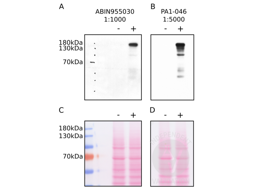
- ABIN955030 reveals a predominant protein band of the expected molecular weight of murine SynGAP in lysates HEK293TF transiently expressing the protein.
Validation #100923 (Western Blotting)
 Validation Images
![HEK293TF cell transfected with a SynGAP expressing (+) or empty pcDNA3 plasmid (-) were lysed and extracted proteins separated in duplicates on a poly-acrylamine gel and transferred to nitrocellulose membrane. Separated proteins were incubated either with the anti-SYNGAP1 antibody ABIN955030 (A) or a different anti-SynGAP antibody (B) at the indicated dilutions. Protein transfer to the nitrocellulose membranes was verified by PonceauS staining prior to the immunoblot (C, D).]() HEK293TF cell transfected with a SynGAP expressing (+) or empty pcDNA3 plasmid (-) were lysed and extracted proteins separated in duplicates on a poly-acrylamine gel and transferred to nitrocellulose membrane. Separated proteins were incubated either with the anti-SYNGAP1 antibody ABIN955030 (A) or a different anti-SynGAP antibody (B) at the indicated dilutions. Protein transfer to the nitrocellulose membranes was verified by PonceauS staining prior to the immunoblot (C, D).
Full Methods
HEK293TF cell transfected with a SynGAP expressing (+) or empty pcDNA3 plasmid (-) were lysed and extracted proteins separated in duplicates on a poly-acrylamine gel and transferred to nitrocellulose membrane. Separated proteins were incubated either with the anti-SYNGAP1 antibody ABIN955030 (A) or a different anti-SynGAP antibody (B) at the indicated dilutions. Protein transfer to the nitrocellulose membranes was verified by PonceauS staining prior to the immunoblot (C, D).
Full Methods -
- Concentration
- 0,5 mg/mL
- Buffer
- Tris saline, pH 7.3 containing 0.02 % sodium azide as preservative and 0.5 % bovine serum albumin as stabilizer
- Preservative
- Sodium azide
- Precaution of Use
- This product contains sodium azide: a POISONOUS AND HAZARDOUS SUBSTANCE which should be handled by trained staff only.
- Handling Advice
- Avoid repeated freezing and thawing.
- Storage
- 4 °C/-20 °C
- Storage Comment
- Store the antibody undiluted at 2-8 °C for one month or (in aliquots) at -20 °C for longer.
-
- Target
- SYNGAP1 (Synaptic Ras GTPase Activating Protein 1 (SYNGAP1))
- Alternative Name
- SYNGAP1 (SYNGAP1 Products)
- Synonyms
- SYNGAP1 antibody, Syngap antibody, MRD5 antibody, RASA1 antibody, RASA5 antibody, SYNGAP antibody, Gm1963 antibody, synaptic Ras GTPase activating protein 1 antibody, zinc finger and BTB domain containing 9 antibody, synaptic Ras GTPase activating protein 1 homolog (rat) antibody, SYNGAP1 antibody, ZBTB9 antibody, syngap1 antibody, Syngap1 antibody
- Background
- Synonyms: KIAA1938, Neuronal RasGAP, RASA1, RASA5, Ras GTPase-activating protein SynGAP, SYNGAP, Synaptic Ras-GAP 1
- Gene ID
- 8831
- NCBI Accession
- NP_006763
- Pathways
- Regulation of long-term Neuronal Synaptic Plasticity
-


