Retinoblastoma 1 antibody (pSer795)

 (1 validation)
Quick Overview for Retinoblastoma 1 antibody (pSer795) (ABIN712946)
Target
See all Retinoblastoma 1 (RB1) AntibodiesReactivity
Host
Clonality
Conjugate
Application
-
-
Binding Specificity
- pSer795
-
Predicted Reactivity
- Human,Mouse,Rat,Dog,Cow,Horse,Rabbit
-
Purification
- Purified by Protein A.
-
Immunogen
- KLH conjugated synthetic phosphopeptide derived from human P105 RB around the phosphorylation site of Ser795
-
Isotype
- IgG
-
-
anti-Retinoblastoma 1 (RB1) (pSer780) antibody
RB1 Reactivity: Human, Mouse, Rat WB, IHC, ELISA, IF, ICC Host: Rabbit Polyclonal unconjugated
anti-Retinoblastoma 1 (RB1) (pSer807) antibodyRB1 Reactivity: Human, Mouse, Rat WB, IHC, ELISA, IF, ICC Host: Rabbit Polyclonal unconjugated
anti-Retinoblastoma 1 (RB1) (pSer795) antibodyRB1 Reactivity: Human, Mouse, Rat WB, IHC, ELISA, IF, ICC Host: Rabbit Polyclonal unconjugated
anti-Retinoblastoma 1 (RB1) (C-Term) antibodyRB1 Reactivity: Human, Mouse, Rat WB, IHC, ELISA, IF, ICC Host: Rabbit Polyclonal unconjugated
anti-Retinoblastoma 1 (RB1) (pSer780) antibodyRB1 Reactivity: Human WB, IHC Host: Rabbit Monoclonal unconjugated
anti-Retinoblastoma 1 (RB1) (C-Term) antibodyRB1 Reactivity: Human WB, IF, IP, IHC (p), ICC Host: Rabbit Polyclonal unconjugated
anti-Retinoblastoma 1 (RB1) (pSer612) antibodyRB1 Reactivity: Human WB, IHC (p) Host: Rabbit Polyclonal RB7659 unconjugated
anti-Retinoblastoma 1 (RB1) (AA 586-615) antibodyRB1 Reactivity: Human WB, IF, IHC (p) Host: Rabbit Polyclonal RB7977 unconjugated
anti-Retinoblastoma 1 (RB1) (AA 858-886), (C-Term) antibodyRB1 Reactivity: Human WB, IHC (p), FACS Host: Rabbit Polyclonal RB20715 unconjugated
-
-
Application Notes
-
WB 1:300-5000
ELISA 1:500-1000
IHC-P 1:200-400
IHC-F 1:100-500
IF(IHC-P) 1:50-200
IF(IHC-F) 1:50-200
IF(ICC) 1:50-200 -
Restrictions
- For Research Use only
-
-
- by
- Kinexus Bioinformatics Corporation
- No.
- #029821
- Date
- 09/21/2014
- Antigen
- Lot Number
- 121026
- Method validated
- Western Blotting
- Positive Control
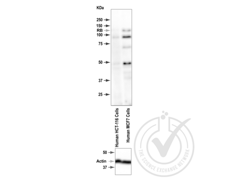
- MCF7 cells
- Negative Control
- HCT-116 cells - low
- Notes
- A band was observed in the positive control at the expected size (~100-105 kDa), which was absent from the negative control. There were some additional bands observed at non-expected molecular weights in the positive control.
- Primary Antibody
- Antigen: Retinoblastoma 1 (RB1) (pSer795)
- Catalog number: ABIN712946
- Lot number: 121026
- Dilution: 1:100
- Secondary Antibody
- Antibody: Donkey anti-Rabbit IgG Antibody (HRP)
- Lot number: F0613
- Dilution: 1:10,000
- Full Protocol
- 1. Cell/tissue total protein lysates were boiled in 1X SDS Sample Buffer containing 1% SDS and 1.25% β-mercaptoethanol at 95°C for 5 minutes prior to loading.
- 2. 15 μg of boiled lysate were loaded and resolved on a 12% SDS-polyacrylamide gel.
- 3. The Precision Plus Protein™ All Blue Prestained Standards from BioRad (161-0373) were used as molecular mass markers.
- 4. Proteins were transferred onto nitrocellulose membrane by tank transfer and protein transfer was confirmed with Ponceau S staining.
- 5. The immunoblot membrane was blocked in 2.5% skim milk and 1.5% BSA solution in TTBS at room temperature for 60 minutes.
- 6. The membrane was washed in TTBS twice for 5 minutes each.
- 7. The membrane was immersed with the protein side up in the antibody solution in TBS and incubated overnight at 4°C with gentle agitation.
- 8. The membrane was rinsed twice with TTBS.
- 9. The membrane was washed in TTBS twice for 5 minutes each.
- 10. The membrane was washed in TTBS once for 15 minutes.
- 11. The membrane was incubated in the HRP-conjugated secondary antibody solution in TBS for 60 minutes at room temperature with gentle agitation.
- 12. The membrane was rinsed twice with TTBS.
- 13. The membrane was washed in TTBS twice for 5 minutes each.
- 14. The membrane was washed in TTBS once for 15 minutes.
- 15. Signals were detected by chemiluminescence (ECL). The blot was scanned for 320 seconds.
- 16. The membrane was rinsed three times with TTBS.
- 17. Repeated Steps 4-15 with the loading control antibody and its matching secondary antibody. The blot was scanned for 160 seconds.
- Experimental Notes
- - No experimental challenges noted.
Validation #029821 (Western Blotting)
 Validation Images
![Figure 1: Western Blot for RB1 (pSer795). Grey arrowhead indicates the expected molecular weight of ~100-105 kDa.]() Figure 1: Western Blot for RB1 (pSer795). Grey arrowhead indicates the expected molecular weight of ~100-105 kDa.
Full Methods
Figure 1: Western Blot for RB1 (pSer795). Grey arrowhead indicates the expected molecular weight of ~100-105 kDa.
Full Methods -
-
Format
- Liquid
-
Concentration
- 1 μg/μL
-
Buffer
- 0.01M TBS( pH 7.4) with 1 % BSA, 0.02 % Proclin300 and 50 % Glycerol.
-
Preservative
- ProClin
-
Precaution of Use
- This product contains ProClin: a POISONOUS AND HAZARDOUS SUBSTANCE, which should be handled by trained staff only.
-
Storage
- 4 °C,-20 °C
-
Storage Comment
- Shipped at 4°C. Store at -20°C for one year. Avoid repeated freeze/thaw cycles.
-
Expiry Date
- 12 months
-
-
- Retinoblastoma 1 (RB1)
-
Alternative Name
- P105 RB
-
Background
-
Synonyms: RbSer795, RB1phospho S795, OSRC, P105 RB, P105RB, PP105, PP110, pRb, RB 1, RB1, RB1 protein, Retinoblastoma 1 including osteosarcoma, Retinoblastoma 1, Retinoblastoma associated protein, Retinoblastoma related osteosarcoma, Retinoblastoma susceptibility gene, Including osteosarcoma, RB_HUMAN.
Background: Rb is a tumor suppressor gene which functions as a negative regulator of the cell cycle by interacting with transcription factors including E2F1, PU1, ATF2, UBF, Elf1 and cAbl. This ability of Rb to alter transcription is regulated by phosphorylation catalyzed by the cyclin dependent protein kinases (cdks). Rb is phosphorylated on serine and threonine, but not on tyrosine residues. It forms a complex with SV40 large T antigen, adenovirus E1A, and human papilloma virus 16E. Rb protein may act by regulating transcription and loss of its function leads to uncontrolled cell growth. Aberrations in the Rb gene have been implicated in cancers of breast, colon, prostate, kidney, nasopharynx, and leukemia.
-
Gene ID
- 5925
-
Pathways
- Cell Division Cycle, Intracellular Steroid Hormone Receptor Signaling Pathway, Mitotic G1-G1/S Phases, DNA Replication, Maintenance of Protein Location, Synthesis of DNA, Autophagy
Target
-