Retinoblastoma 1 antibody (pSer780)
 (1 reference)
 (1 validation)
Quick Overview for Retinoblastoma 1 antibody (pSer780) (ABIN675294)
Target
See all Retinoblastoma 1 (RB1) AntibodiesReactivity
Host
Clonality
Conjugate
Application
-
-
Binding Specificity
- pSer780
-
Cross-Reactivity
- Human, Rat
-
Predicted Reactivity
- Mouse,Dog,Cow,Chicken
-
Purification
- Purified by Protein A.
-
Immunogen
- KLH conjugated synthetic phosphopeptide derived from human Rb around the phosphorylation site of (Ser780)
-
Isotype
- IgG
-
-
anti-Retinoblastoma 1 (RB1) (pSer780) antibody
RB1 Reactivity: Human WB, IHC Host: Rabbit Monoclonal unconjugated
anti-Retinoblastoma 1 (RB1) (pSer780) antibodyRB1 Reactivity: Human, Mouse, Rat WB, IHC, ELISA, IF, ICC Host: Rabbit Polyclonal unconjugated
anti-Retinoblastoma 1 (RB1) (pSer807) antibodyRB1 Reactivity: Human, Mouse, Rat WB, IHC, ELISA, IF, ICC Host: Rabbit Polyclonal unconjugated
anti-Retinoblastoma 1 (RB1) (pSer795) antibodyRB1 Reactivity: Human, Mouse, Rat WB, IHC, ELISA, IF, ICC Host: Rabbit Polyclonal unconjugated
anti-Retinoblastoma 1 (RB1) (C-Term) antibodyRB1 Reactivity: Human, Mouse, Rat WB, IHC, ELISA, IF, ICC Host: Rabbit Polyclonal unconjugated
anti-Retinoblastoma 1 (RB1) (AA 586-615) antibodyRB1 Reactivity: Human WB, IF, IHC (p) Host: Rabbit Polyclonal RB7977 unconjugated
anti-Retinoblastoma 1 (RB1) (pSer612) antibodyRB1 Reactivity: Human WB, IHC (p) Host: Rabbit Polyclonal RB7659 unconjugated
anti-Retinoblastoma 1 (RB1) (AA 858-886), (C-Term) antibodyRB1 Reactivity: Human WB, IHC (p), FACS Host: Rabbit Polyclonal RB20715 unconjugated
anti-Retinoblastoma 1 (RB1) (C-Term) antibodyRB1 Reactivity: Human WB, IF, IP, IHC (p), ICC Host: Rabbit Polyclonal unconjugated
-
-
Application Notes
-
WB 1:300-5000
ELISA 1:500-1000
FCM 1:20-100
IHC-P 1:200-400
IHC-F 1:100-500
IF(IHC-P) 1:50-200
IF(IHC-F) 1:50-200
IF(ICC) 1:50-200 -
Restrictions
- For Research Use only
-
-
- by
- Alamo Laboratories Inc
- No.
- #029795
- Date
- 08/21/2014
- Antigen
- Retinoblastoma 1 (RB1) (pSer780)
- Lot Number
- 120508
- Method validated
- Western Blotting
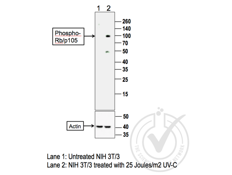
- Positive Control
- NIH/3T3 cells irradiated with 25 Joules/m2 UV-C
- Negative Control
- Non-irradiated NIH/3T3 cells
- Notes
- A strong band was observed in the positive control sample at the correct molecular weight. An additional band was also observed in the positive sample at a lower molecular weight which may represent non-specific binding. No bands were observed in the negative control sample.
- Primary Antibody
- Antigen: Retinoblastoma 1 (RB1) (pSer780)
- Catalog number: ABIN675294
- Lot number: 120508
- Antibody Dilution: 1:100
- Secondary Antibody
- Antigen: Goat Anti-Rabbit IgG (H + L)-HRP Conjugate
- Lot number: L170-6515
- Antibody Dilution: 1:10,000
- Full Protocol
- 1. The cell extracts were heated at 95°C for 5 minutes in 1X SDS Sample Buffer containing 1% SDS and 1.25% β-mercaptoethanol.
- 2. 20 μL of heated culture-media were loaded and resolved on 8-16% SDS-polyacrylamide gel.
- 3. The Thermo Scientific - Spectra Multicolor Broad Range (Cat # 26634) were used as molecular mass markers.
- 4. Proteins were then transferred onto PVDF membrane by wet transfer and protein transfer was confirmed with Ponceau-S staining.
- 5. The PVDF membrane was incubated with 25 mL of blocking buffer [Tris Buffered Saline, pH 7.4 plus 0.1% TW20 (TBST)] containing 5% (W/V) BSA at room temperature for 1 hour.
- 6. The membrane was rinsed with TBST once.
- 7. The membrane was immersed with the protein side up in the primary antibody solution in TBST containing 5% (W/V) BSA and incubated for 20 hours at 4°C.
- 8. The membrane was rinsed in TBST thrice for 5 minutes each.
- 9. The membrane was incubated in the HRP-conjugated secondary antibody solution in TBST containing 5% (W/V) BSA and incubated for 1 hour at room temperature (~26°C) with gentle agitation.
- 10. The membrane was rinsed thrice TBST thrice for 5 minutes each.
- 11. The membrane was rinsed in TBS twice for 30 seconds each.
- 12. Signals were detected with ECL-2 Substrate. The blot was scanned for 50 minutes.
- 13. The membrane was rinsed three times TBST.
- 14. Incubated in Acidic Glycine Stripping Buffer at room temperature with gentle agitation for 3 times, 10 minutes each.
- 15. The membrane was washed in TBST 2 times for 10 minutes each.
- 16. Repeated Steps 5-12 with the loading control antibody (for Anti-actin) and its matching secondary antibody.
- Experimental Notes
- - No experimental challenges noted.
Validation #029795 (Western Blotting)
 Validation Images
![Figure 1: Western Blot for RB1 pSer780. Grey arrowhead indicates the expected molecular weight of ~105 kDa.]() Figure 1: Western Blot for RB1 pSer780. Grey arrowhead indicates the expected molecular weight of ~105 kDa.
Full Methods
Figure 1: Western Blot for RB1 pSer780. Grey arrowhead indicates the expected molecular weight of ~105 kDa.
Full Methods -
-
Format
- Liquid
-
Concentration
- 1 μg/μL
-
Buffer
- 0.01M TBS( pH 7.4) with 1 % BSA, 0.02 % Proclin300 and 50 % Glycerol.
-
Preservative
- ProClin
-
Precaution of Use
- This product contains ProClin: a POISONOUS AND HAZARDOUS SUBSTANCE, which should be handled by trained staff only.
-
Storage
- 4 °C,-20 °C
-
Storage Comment
- Shipped at 4°C. Store at -20°C for one year. Avoid repeated freeze/thaw cycles.
-
Expiry Date
- 12 months
-
-
-
: "EZH2 enables germinal centre formation through epigenetic silencing of CDKN1A and an Rb-E2F1 feedback loop." in: Nature communications, Vol. 8, Issue 1, pp. 877, (2018) (PubMed).
-
: "EZH2 enables germinal centre formation through epigenetic silencing of CDKN1A and an Rb-E2F1 feedback loop." in: Nature communications, Vol. 8, Issue 1, pp. 877, (2018) (PubMed).
-
- Retinoblastoma 1 (RB1)
-
Alternative Name
- Rb/p105-Rb
-
Background
-
Synonyms: RbSer780, OSRC, P105 RB, P105RB, PP105, PP110, pRb, RB 1, RB1, RB1 protein, Retinoblastoma 1 including osteosarcoma, Retinoblastoma 1, Retinoblastoma associated protein, Including osteosarcoma, Osteosarcoma, p105-Rb, Rb, RB_HUMAN, Retinoblastoma suspectibility protein, Retinoblastoma-associated protein, Retinoblastoma related osteosarcoma, Retinoblastoma susceptibility gene.
Background: Nuclear Marker.The protein encoded by this gene is a negative regulator of the cell cycle and was the first tumor suppressor gene found. The encoded protein also stabilizes constitutive heterochromatin to maintain the overall chromatin structure. The active, hypophosphorylated form of the protein binds transcription factor E2F1. Defects in this gene are a cause of childhood cancer retinoblastoma (RB), bladder cancer, and osteogenic sarcoma.
-
Gene ID
- 5925
-
Pathways
- Cell Division Cycle, Intracellular Steroid Hormone Receptor Signaling Pathway, Mitotic G1-G1/S Phases, DNA Replication, Maintenance of Protein Location, Synthesis of DNA, Autophagy
Target
-


