TCF7L1 antibody (N-Term)
 (1 reference)
 (1 validation)
Quick Overview for TCF7L1 antibody (N-Term) (ABIN6972849)
Target
See all TCF7L1 AntibodiesReactivity
Host
Clonality
Conjugate
Application
-
-
Binding Specificity
- N-Term
-
Characteristics
- TCF3 / TCF7L1 (T-cell factor 3, TCF7L1) is a member of the TCF/LEF family and a component of the Wnt signaling pathway and a dominant downstream effector in embryonic stem cells (ESCs). TCF3 binds to DNA and serves as both a repressor as well as an activator of transcription. TCF3 brings developmental signals directly to the core regulatory circuitry of ES cells to influence the balance between pluripotency and differentiation. TCF3 transcriptionally represses many genes important for maintaining pluripotency and self-renewal, as well as those involved in lineage commitment and stem cell differentiation. This effect is in part mediated by the corepressors transducin-like enhancer of split 2 (TLE2) and C-terminal Binding Protein (CtBP). TCF7L1 / TCF3 antibody (pAb) was raised in a Rabbit host. It has been validated for use in Chromatin Immunoprecipitation, ChIP-Seq and Western blot, it has been shown to react with Human samples.
-
Purification
- Affinity Purified
-
Immunogen
- This TCF7L1 / TCF3 antibody was raised against a peptide from the N-terminus of human TCF7L1 / TCF3.
-
Isotype
- IgG
-
-
anti-Transcription Factor 7-Like 1 (T-Cell Specific, HMG-Box) (TCF7L1) antibody
KD Validated TCF7L1 Reactivity: Human WB, FACS Host: Rabbit Monoclonal 24GB1985 unconjugated Recombinant Antibody
anti-Transcription Factor 7-Like 1 (T-Cell Specific, HMG-Box) (TCF7L1) (AA 35-84) antibodyTCF7L1 Reactivity: Human, Mouse, Rat, Cow, Rabbit, Guinea Pig WB, IHC, IHC (p) Host: Rabbit Polyclonal unconjugated
anti-Transcription Factor 7-Like 1 (T-Cell Specific, HMG-Box) (TCF7L1) (Internal Region) antibodyTCF7L1 Reactivity: Human, Mouse, Rat WB, ELISA, ICC, IF Host: Rabbit Polyclonal unconjugated
anti-Transcription Factor 7-Like 1 (T-Cell Specific, HMG-Box) (TCF7L1) (AA 489-586) antibodyTCF7L1 Reactivity: Human WB, ELISA Host: Mouse Polyclonal unconjugated
anti-Transcription Factor 7-Like 1 (T-Cell Specific, HMG-Box) (TCF7L1) (AA 561-588), (C-Term) antibodyTCF7L1 Reactivity: Human WB, IHC Host: Rabbit Polyclonal unconjugated
anti-Transcription Factor 7-Like 1 (T-Cell Specific, HMG-Box) (TCF7L1) (AA 70-98), (N-Term) antibodyTCF7L1 Reactivity: Human WB Host: Rabbit Polyclonal RB43616 unconjugated
anti-Transcription Factor 7-Like 1 (T-Cell Specific, HMG-Box) (TCF7L1) (AA 80-99) antibodyTCF7L1 Reactivity: Human WB, ELISA Host: Rabbit Polyclonal unconjugated
anti-Transcription Factor 7-Like 1 (T-Cell Specific, HMG-Box) (TCF7L1) (AA 72-121) antibodyTCF7L1 Reactivity: Human, Mouse, Rabbit WB Host: Rabbit Polyclonal unconjugated
anti-Transcription Factor 7-Like 1 (T-Cell Specific, HMG-Box) (TCF7L1) (N-Term) antibodyTCF7L1 Reactivity: Human, Mouse, Rat, Cow, Dog, Guinea Pig WB, IHC Host: Rabbit Polyclonal unconjugated
-
-
Application Notes
-
The rabbit anti-TC7L1 antibody ABIN6972849 is suitable for use in CUT&RUN, ChIP-seq, ChIP, and Western Blot. Specific conditions for each assay should be optimized by the end user. General dilution recommendations for different applications are as follows:
ChIP: 10µL per ChIP
ChIP-seq: 10µL per ChIP
WB: 1:500-1:2,000
CUT&RUN: 1:100 -
Restrictions
- For Research Use only
-
-
- by
- Gianluca Zambanini, Anna Nordin and Claudio Cantù; Cantù Lab, Gene Regulation during Development and Disease, Linköping University
- No.
- #104351
- Date
- 02/28/2022
- Antigen
- TCF7L1
- Lot Number
- 20011001
- Method validated
- Cleavage Under Targets and Release Using Nuclease
- Positive Control
Recombinant anti-H3K27me3 CUT&RUN Positive Control antibody (antibodies-online, ABIN6923144)
- Negative Control
Polyclonal guinea Pig anti-rabbit IgG (antibodies-online, ABIN101961)
- Notes
Passed. ABIN6972849 allows for TCF7L1 targeted digestion using CUT&RUN in human HEK293T cells.
- Primary Antibody
- ABIN6972849
- Secondary Antibody
- Full Protocol
- Cell harvest and nuclear extraction
- Harvest 250,000 HEK293T cells per antibody to be used at RT stimulated with 10 µM CHIR for 24 h at RT.
- Centrifuge cell solution 5 min at 600 x g at RT.
- Remove the liquid carefully.
- Gently resuspend cells in 1 mL of Nuclear Extraction Buffer (20 mM HEPES-KOH pH 8.2, 20% Glycerol, 0,05% IGEPAL, 0.5 mM Spermidine, 10 mM KCl, Roche Complete Protease Inhibitor EDTA-free).
- Move the solution to a 2 mL centrifuge tube.
- Pellet the nuclei 800 x g for 5 min.
- Repeat the NE wash twice for a total of three washes.
- Resuspend the nuclei in 20 µL NE Buffer per sample.
- Concanavalin A beads preparation
- Prepare one 2 mL microcentrifuge tube.
- Gently resuspend the magnetic Concanavalin A Beads (antibodies-online, ABIN6952467).
- Pipette 20 µL Con A Beads slurry for each sample into the 2 mL microcentrifuge tube.
- Place the tube on a magnet stand until the fluid is clear. Remove the liquid carefully.
- Remove the microcentrifuge tube from the magnetic stand.
- Pipette 1 mL Binding Buffer (20 mM HEPES pH 7.5, 10 mM KCl, 1 mM CaCl2, 1 mM MnCl2) into the tube and resuspend ConA beads by gentle pipetting.
- Spin down the liquid from the lid with a quick pulse in a table-top centrifuge.
- Place the tubes on a magnet stand until the fluid is clear. Remove the liquid carefully.
- Remove the microcentrifuge tube from the magnetic stand.
- Repeat the wash twice for a total of three washes.
- Gently resuspend the ConA Beads in a volume of Binding Buffer corresponding to the original volume of bead slurry, i.e. 20 µL per sample.
- Nuclei immobilization – binding to Concanavalin A beads
- Carefully vortex the nuclei suspension and add 20 µL of the Con A beads in Binding Buffer to the cell suspension for each sample.
- Close tube tightly incubates 10 min at 4 °C.
- Put the 2 mL tube on the magnet stand and when the liquid is clear remove the supernatant.
- Resuspend the beads in 1 mL of EDTA Wash buffer (20 mM HEPES pH 7.5, 150 mM NaCl, 0.5 mM Spermidine, Roche Complete Protease Inhibitor EDTA-free, 2mM EDTA).
- Incubate 5 min at RT.
- Place the tube on the magnet stand and when the liquid is clear remove the supernatant.
- Resuspend the beads in 200 µl of Wash Buffer (20 mM HEPES pH 7.5, 150 mM NaCl, 0.5 mM Spermidine, Roche Complete Protease Inhibitor EDTA-free) per sample.
- Primary antibody binding
- Divide nuclei suspension into separate 200 µL PCR tubes, one for each antibody.
- Add 2 µL antibody (anti-TCF7L1 antibody ABIN6972849, anti-H3K27me3 antibody positive control ABIN6923144, and guinea pig anti-rabbit IgG negative control antibody ABIN101961) to the respective tube, corresponding to a 1:100 dilution.
- Incubate at 4 °C ON.
- Place the tubes on a magnet stand until the fluid is clear. Remove the liquid carefully.
- Remove the microcentrifuge tubes from the magnetic stand.
- Wash with 200 µL of Wash Buffer using a multichannel pipette to accelerate the process.
- Repeat the wash five times for a total of six washes.
- pAG-MNase Binding
- Prepare a 1.5 mL microcentrifuge tube containing 100 µL of pAG mix per sample (100 µL of wash buffer + 58.5 µg pAG-MNase per sample).
- Place the PCR tubes with the sample on a magnet stand until the fluid is clear. Remove the liquid carefully.
- Remove tubes from the magnetic stand.
- Resuspend the beads in 100 µL of pAG-MNase premix.
- Incubate 30 min at 4 °C.
- Place the tubes on a magnet stand until the fluid is clear. Remove the liquid carefully.
- Remove the microcentrifuge tubes from the magnetic stand.
- Wash with 200 µL of Wash Buffer using a multichannel pipette to accelerate the process.
- Repeat the wash five times for a total of six washes.
- Resuspend in 100 µL of Wash Buffer.
- MNase digestion and release of pAG-MNase-antibody-chromatin complexes
- Place PCR tubes on ice and allow to chill.
- Prepare a 1.5 mL microcentrifuge tube with 102 µl of 2 mM CaCl2 mix per sample (100 µl Wash Buffer + 2 µL 100 mM CaCl2) and let it chill on ice.
- Always in ice, place the samples on the magnetic rack and when the liquid is clear remove the supernatant.
- Resuspend the samples in 100 µl of the 2 mM CaCl2 mix and incubate in ice for exactly 30 min.
- Place the sample on the magnet stand and when the liquid is clear remove the supernatant.
- Resuspend the sample in 50 µl of 1x Urea STOP Buffer (8.5 M Urea, 100 mM NaCl, 2 mM EGTA, 2 mM EDTA, 0,5% IGEPAL).
- Incubate the samples 1h at 4°C.
- Transfer the supernatant containing the pAG-MNase-bound digested chromatin fragments to fresh 200 µl PCR tubes.
- DNA Clean up
- Take the Mag-Bind® TotalPure NGS beads (Omega Bio-Tek, M1378-01) from the storage and wait until they are at RT.
- Add 2x volume of beads to each sample (e.g. 100 µL of beads for 50 µL of sample).
- Incubate the beads and the sample for 15 min at RT.
- During incubation prepare fresh EtOH 80%.
- Place the PCR tubes on a magnet stand and when the liquid is clear remove the supernatant.
- Add 200 µl of fresh 80% EtOH to the sample without disturbing the beads (Important!!! Do NOT resuspend the beads or remove the tubes from the magnet stand or the sample will be lost).
- Incubate 30 sec at RT.
- Remove the EtOH from the sample.
- Repeat the wash with 80% EtOH.
- Resuspend the beads in 25 µL of 10 mM Tris.
- Incubate the sample for 2 min at RT.
- Repeat the 2x beads clean up as described before (this time with 50 µL of beads for each sample).
- Resuspend the beads + DNA in 20 µL of 10 mM Tris.
- Library preparation and sequencing
- Prepare Libraries using KAPA HyperPrep Kit using KAPA Dual-Indexed adapters according to protocol.
- Sequence samples on an Illumina NextSeq 500 sequencer, using a NextSeq 500/550 High Output Kit v2.5 (75 Cycles), 36 bp PE.
- Peak calling
- Trim reads using using bbTools bbduk (BBMap - Bushnell B. - sourceforge.net/projects/bbmap/) to remove adapters, artifacts and repeat sequences.
- Map aligned reads to the hg38 human genome using bowtie with options -m 1 -v 0 -I 0 -X 500.
- Use SAMtools to convert SAM files to BAM files and remove duplicates.
- Use BEDtools genomecov to produce Bedgraph files.
- Call peaks using SEACR with a 0.001 threshold and the option norm stringent.
- Experimental Notes
Results are published in Zambanini, G. et al. A New CUT&RUN Low Volume-Urea (LoV-U) protocol uncovers Wnt/β-catenin tissue-specific genomic targets. bioRxiv (2022). https://doi.org/10.1101/2022.07.06.498999
Validation #104351 (Cleavage Under Targets and Release Using Nuclease)
 Validation Images
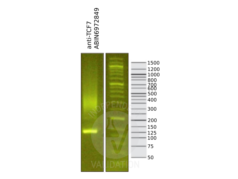
![Library profiles comparing fragment size distributions on an E-Gel EX 2% agarose gel (Thermo Fisher). Fragments obtained from CUT&RUN using anti-TCF7L1 antibody ABIN6972849 after library preparation, compared to the E-Gel Sizing DNA Ladder (Thermo Fisher).]() Library profiles comparing fragment size distributions on an E-Gel EX 2% agarose gel (Thermo Fisher). Fragments obtained from CUT&RUN using anti-TCF7L1 antibody ABIN6972849 after library preparation, compared to the E-Gel Sizing DNA Ladder (Thermo Fisher).
Library profiles comparing fragment size distributions on an E-Gel EX 2% agarose gel (Thermo Fisher). Fragments obtained from CUT&RUN using anti-TCF7L1 antibody ABIN6972849 after library preparation, compared to the E-Gel Sizing DNA Ladder (Thermo Fisher).
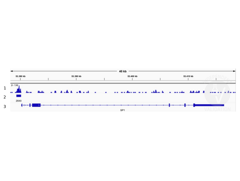
![1. Alignment tracks from CUT&RUN targeting TCF7L1 in HEK293T cells using anti-TCF7L1 antibody ABIN6972849. 2. Peaks called by SEACR from CUT&RUN data using anti-TCF7L1 antibody ABIN6972849. 3. RefSeq Genes.]() 1. Alignment tracks from CUT&RUN targeting TCF7L1 in HEK293T cells using anti-TCF7L1 antibody ABIN6972849. 2. Peaks called by SEACR from CUT&RUN data using anti-TCF7L1 antibody ABIN6972849. 3. RefSeq Genes.
Full Methods
1. Alignment tracks from CUT&RUN targeting TCF7L1 in HEK293T cells using anti-TCF7L1 antibody ABIN6972849. 2. Peaks called by SEACR from CUT&RUN data using anti-TCF7L1 antibody ABIN6972849. 3. RefSeq Genes.
Full Methods -
-
Buffer
- Purified IgG in PBS with 30 % glycerol and 0.035 % sodium azide.
-
Preservative
- Sodium azide
-
Precaution of Use
- This product contains Sodium azide: a POISONOUS AND HAZARDOUS SUBSTANCE which should be handled by trained staff only.
-
Storage
- -20 °C
-
Storage Comment
- Avoid repeated freeze/thaw cycles by aliquoting items into single-use fractions for storage at -20°C for up to 2 years. Keep all reagents on ice when not in storage.
-
-
-
: "A new cut&run low volume-urea (LoV-U) protocol optimized for transcriptional co-factors uncovers Wnt/b-catenin tissue-specific genomic targets." in: Development (Cambridge, England), (2022) (PubMed).
-
-
- TCF7L1 (Transcription Factor 7-Like 1 (T-Cell Specific, HMG-Box) (TCF7L1))
-
Alternative Name
- TCF7L1 / TCF3
-
Molecular Weight
- 80 kDa
-
NCBI Accession
- NP_112573
-
Pathways
- WNT Signaling, Stem Cell Maintenance
Target
-


