MEK1 Antibodies
Your search for reliable MEK1 antibodies ends here. MEK1, known by aliases such as Map2k1, MAP2K1, map2k1.L, map2k1, MEK1, is an integral part of our antibody range. Whether you're working with Human, Rat, Mouse, Dog, Cow, or other species, our range of MEK1 antibodies offer precise detection across diverse samples. These specialized antibodies are tailored for various scientific applications like WB, ELISA, IHC, IF (cc), IF (p), providing you with options like polyclonal, recombinant, and monoclonal antibodies, sourced from different host species such as Rabbit, Mouse, Chicken. The efficacy of our antibodies is well-established, demonstrated through multiple methods.
Detailed information, including references, images, and validations by other customers, can be found on each product page. Should you require assistance in finding a specific product, our customer service team is ready to assist. Utilize our MEK1 antibodies in your research endeavors for dependable MEK1 detection.
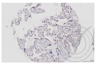
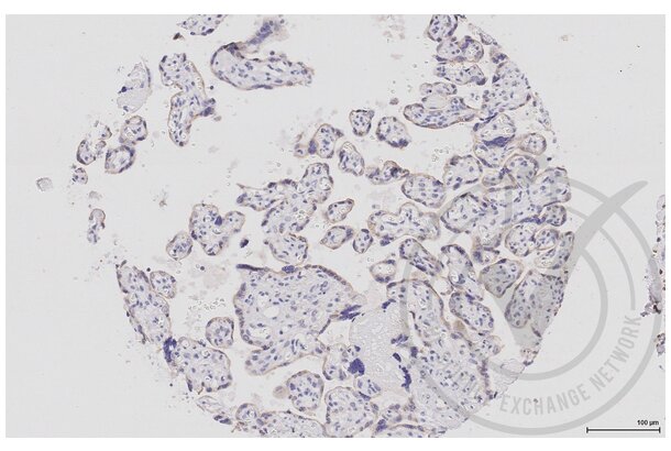 MEK1 antibody (AA 2-150) (ABIN686482)
MAP2K1 Reactivity: Human ELISA, IHC (p), IF (cc), IF (p), FACS, IHC (fro) Host: Rabbit Polyclonal unconjugated
MAP2K1 Reactivity: Human, Mouse, Rat, Dog, Monkey WB, IHC, IF, IHC (p) Host: Mouse Monoclonal 2H3 unconjugated
MAP2K1 Reactivity: Human, Mouse, Rat, Dog, Monkey WB, IHC, IF, IHC (p) Host: Mouse Monoclonal 3H3 unconjugated
MEK1 Antibodies by Host
Find MEK1 Antibodies with a specific Host. The Host listed below are among those available. Click on a link to go to the corresponding products.
MEK1 Antibodies by Clonality
Find available monoclonal or polyclonal MEK1 Antibodies. Click on a link to go to the corresponding products.
Popular MEK1 Antibodies
- (3)
- (1)
- (6)
- (9)
- (1)
- (5)
- (5)
- (6)
- (4)
- (4)
- (3)
- (3)
- (3)
- (2)
- (2)
- (3)
- (3)
- (3)
Latest Publications for our MEK1 Antibodies
: "Integrated Proteomic and Transcriptomic Analysis Reveals Long Noncoding RNA HOX Transcript Antisense Intergenic RNA (HOTAIR) Promotes Hepatocellular Carcinoma Cell Proliferation by Regulating Opioid ..." in: Molecular & cellular proteomics : MCP, Vol. 17, Issue 1, pp. 146-159, (2019) (PubMed).: "Follicular fluid meiosis-activating sterol (FF-MAS) promotes meiotic resumption via the MAPK pathway in porcine oocytes." in: Theriogenology, (2019) (PubMed).
: "Traditional Chinese Medicine CFF-1 induced cell growth inhibition, autophagy, and apoptosis via inhibiting EGFR-related pathways in prostate cancer." in: Cancer medicine, Vol. 7, Issue 4, pp. 1546-1559, (2018) (PubMed).
: "EGFR‑associated pathways involved in traditional Chinese medicine (TCM)‑1‑induced cell growth inhibition, autophagy and apoptosis in prostate cancer." in: Molecular medicine reports, Vol. 17, Issue 6, pp. 7875-7885, (2018) (PubMed).
: "Cinobufacini inhibits epithelial-mesenchymal transition of human hepatocellular carcinoma cells through c-Met/ERK signaling pathway." in: Bioscience trends, Vol. 12, Issue 3, pp. 291-297, (2018) (PubMed).
: "MCM6 promotes metastasis of hepatocellular carcinoma via MEK/ERK pathway and serves as a novel serum biomarker for early recurrence." in: Journal of experimental & clinical cancer research : CR, Vol. 37, Issue 1, pp. 10, (2018) (PubMed).
: "Induction of Apoptosis in Human Leukemic Cell Lines by Diallyl Disulfide via Modulation of EGFR/ERK/PKM2 Signaling Pathways." in: Asian Pacific journal of cancer prevention : APJCP, Vol. 16, Issue 8, pp. 3509-15, (2015) (PubMed).
: "MiR-203 down-regulates Rap1A and suppresses cell proliferation, adhesion and invasion in prostate cancer." in: Journal of experimental & clinical cancer research : CR, Vol. 34, pp. 8, (2015) (PubMed).
: "Chemotherapy triggers HIF-1-dependent glutathione synthesis and copper chelation that induces the breast cancer stem cell phenotype." in: Proceedings of the National Academy of Sciences of the United States of America, Vol. 112, Issue 33, pp. E4600-9, (2015) (PubMed).
: "MEK1 is associated with carboplatin resistance and is a prognostic biomarker in epithelial ovarian cancer." in: BMC cancer, Vol. 14, pp. 837, (2014) (PubMed).
Aliases for MEK1 Antibodies
mitogen activated protein kinase kinase 1 (Map2k1) Antibodiesmitogen-activated protein kinase kinase 1 (MAP2K1) Antibodies
mitogen-activated protein kinase kinase 1 (Map2k1) Antibodies
mitogen-activated protein kinase kinase 1 L homeolog (map2k1.L) Antibodies
mitogen-activated protein kinase kinase 1 (map2k1) Antibodies
MAP kinase/ ERK kinase 1 (MEK1) Antibodies
ATMEK1 Antibodies
CFC3 Antibodies
F20B18.180 Antibodies
F20B18_180 Antibodies
MAP kinase/ ERK kinase 1 Antibodies
MAPKK1 Antibodies
MEK Antibodies
mek-2 Antibodies
Mek1 Antibodies
MEK1 Antibodies
MEKK1 Antibodies
MITOGEN ACTIVATED PROTEIN KINASE KINASE Antibodies
MITOGEN ACTIVATED PROTEIN KINASE KINASE 1 Antibodies
MKK1 Antibodies
NMAPKK Antibodies
PRKMK1 Antibodies
Prkmk1 Antibodies
si:ch211-242m18.2 Antibodies
wu:fj56a12 Antibodies
wu:fj61b01 Antibodies
zgc:56557 Antibodies
Did you look for something else?
- MEIS3 Antibodies
- MEIS1 Antibodies
- Meis Homeobox 2 Antibodies
- Meiosis 1 Associated Protein Antibodies
- MEIOB Antibodies
- MEIG1 Antibodies
- MEI4 Antibodies
- MEI1 Antibodies
- MEGF8 Antibodies
- MEGF11 Antibodies
- MEGF10 Antibodies
- MEG3 Antibodies
- MEFV Antibodies
- MEF2D Antibodies
- MEF2C Antibodies
- MEF2BNB-MEF2B Readthrough Antibodies
- MEF2BNB Antibodies
- MEF2B Antibodies
- MEF2A Antibodies
- MED9 Antibodies
- Melanocortin 5 Receptor Antibodies
- Melanoma Antigen Family D, 4 Antibodies
- Melanoma Antigen Family D, 4B Antibodies
- Melanophilin Antibodies
- Melatonin Antibodies
- Melatonin Receptor 1A Antibodies
- Melatonin Receptor 1B Antibodies
- MELK Antibodies
- Membrane Protein, Palmitoylated 2 (MAGUK P55 Subfamily Member 2) Antibodies
- Membrane transport protein XK Antibodies
- Membrane-Spanning 4-Domains, Subfamily A, Member 8 Antibodies
- MEMO1 Antibodies
- Menin Antibodies
- Meningioma 1 Antibodies
- MEOX1 Antibodies
- MEOX2 Antibodies
- MEP1A Antibodies
- MEPCE Antibodies
- MEPE Antibodies
- Meprin B Antibodies


